Daniel Wenz1,2 and Rolf Gruetter1,3
1CIBM Center for Biomedical Imaging, Lausanne, Switzerland, 2Animal Imaging and Technology, Ecole Polytechnique Federale de Lausanne (EPFL), Lausanne, Switzerland, 3Laboratory of Functional and Metabolic Imaging (LIFMET), Ecole Polytechnique Federale de Lausanne (EPFL), Lausanne, Switzerland
1CIBM Center for Biomedical Imaging, Lausanne, Switzerland, 2Animal Imaging and Technology, Ecole Polytechnique Federale de Lausanne (EPFL), Lausanne, Switzerland, 3Laboratory of Functional and Metabolic Imaging (LIFMET), Ecole Polytechnique Federale de Lausanne (EPFL), Lausanne, Switzerland
We conclude that the approach presented in this study
has potential to provide new insights into dielectrically-shortened dipole
antenna design and may be particularly relevant given the growing number of
such antenna designs for UHF-MRI.
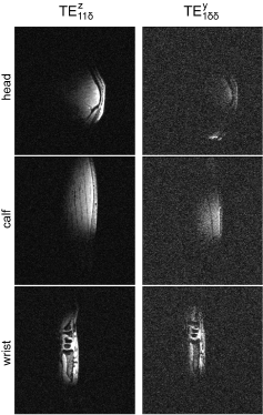
Fig. 5.
In vivo MRI experiments (3D-GRE:
TR/TE = 6.5/2.82ms, FOV = 256x240 mm2, slice thickness = 1.0 mm, FA = 4º,
reference transmit voltage = 100 V) in one human male subject using two blocks:
thinner (d/b = 0.25) and thicker one (d/b = 0.75). Three different regions of
interest (head, calf, wrist) were investigated. The quality of all of the
images was significantly compromised for the larger block (very noisy). The
overall quality of the images depended on the level of curvature of the
anatomical structure. The acquisition parameters of the RF pulse sequence were
used to scan each body part.
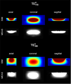
Fig. 4.
Visualization of dielectric modes: the comparison between the electromagnetic
field simulations and MR measurements for two elements: thinner one (d = 0.25b) and thicker one (d =
0.75b). GRE imaging was used
(TR/TE=8.6/4.0 ms, FOV=250 x
250 mm2, slice thickness = 7.0
mm, FA=15º, reference transmit voltage = 5 V). The simulations are in an
excellent agreement with the measurements and show significantly different
magnetic field distribution between the blocks. The mode that propagates within
the thinner block was interpreted as
TE11δz, and within the
thicker one as
TE1δδy.