Moritz Simon Fabian1, Felix Glang2, Katrin Michaela Khakzar1, Angelika Barbara Mennecke1, Alexander German1, Manuel Schmidt3, Burkhard Kasper4, Arnd Dörfler1, Frederik B. Laun1, and Moritz Zaiss1
1Department of Neuroradiology, University Hospital Erlangen, Friedrich Alexander University Erlangen-Nürnberg, Erlangen, Germany, 2High-field Magnetic Resonance Center, Max Planck Institute for Biological Cybernetics, Tübungen, Germany, 3Department of Neuroradiology, University Hospital Erlangen, Erlangen, Germany, 4Department of Neurology, Epilepsy Center, University Hospital Erlangen, Erlangen, Germany
1Department of Neuroradiology, University Hospital Erlangen, Friedrich Alexander University Erlangen-Nürnberg, Erlangen, Germany, 2High-field Magnetic Resonance Center, Max Planck Institute for Biological Cybernetics, Tübungen, Germany, 3Department of Neuroradiology, University Hospital Erlangen, Erlangen, Germany, 4Department of Neurology, Epilepsy Center, University Hospital Erlangen, Erlangen, Germany
The L1-regularized linear
projection approach allows for reduction of scan time by a factor of ~2.8,
while still being able to predict multiple CEST contrast parameters. It
generalizes to unseen healthy subject data and tumor patient data.
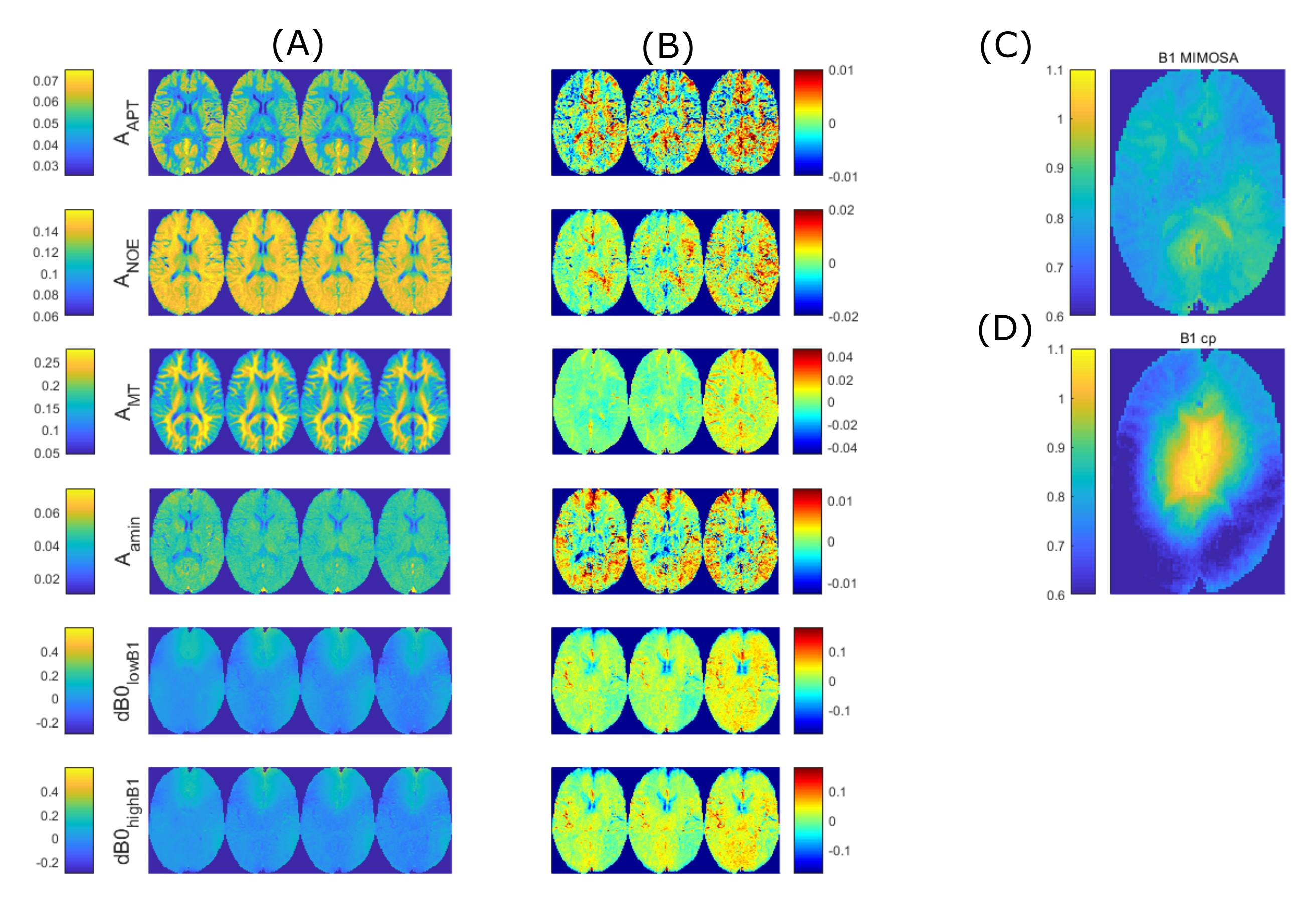
Figure
2: Comparison of retrospective and prospective input feature reduction as well
as conventional Lorentzian fitting. (A)
left to right: ground truth by Lorentzian fitting on full measurement, linear
prediction with all input features contained, linear prediction with retrospective
reduction of input features according to the LASSO-reduced sampling schedule,
measurement with these input features being prospectively omitted. (B) Difference maps of the linear
predictions and the ground truth. (C)
B1-MIMOSA and (D) B1-cp map as mentioned
in 5
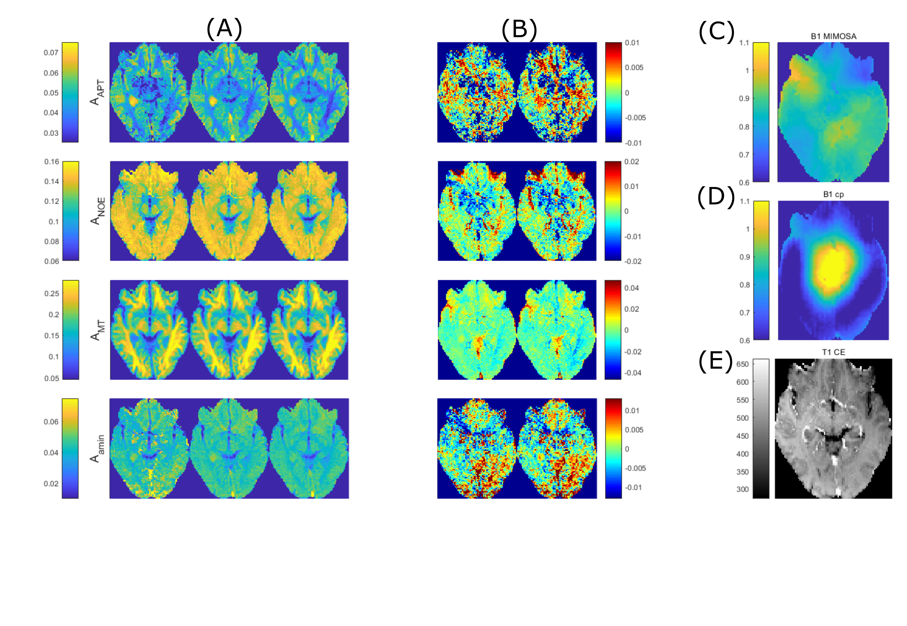
Figure 3: Comparison of
retrospective reduction of input features for a tumor patient dataset
(Glioblastoma WHO grade 4). (A) left
to right: ground truth obtained by Lorentzian fitting, linear prediction with
all input features retained, retrospective reduction of input features. (B) Difference maps between linear
predictions and ground truth. (C) B1
MIMOSA map and (D) B1-cp map as mentioned
in 5. (E) Anatomical T1
contrast-enhanced image