Shu Zhang1, Xinzeng Wang2, F. William Schuler1, R. Marc Lebel3, Mitsuharu Miyoshi4, Ersin Bayram2, Elena Vinogradov5, Jason Michael Johnson6, Jingfei Ma7, and Mark David Pagel1,7
1Cancer Systems Imaging, MD Anderson Cancer Center, Houston, TX, United States, 2Global MR Applications & Workflow, GE Healthcare, Houston, TX, United States, 3Global MR Applications & Workflow, GE Healthcare, Calgary, AB, Canada, 4Global MR Applications & Workflow, GE Healthcare Japan, Tokyo, Japan, 5Radiology, UT Southwestern Medical Center, Dallas, TX, United States, 6Neuroradiology, MD Anderson Cancer Center, Houston, TX, United States, 7Imaging Physics, MD Anderson Cancer Center, Houston, TX, United States
1Cancer Systems Imaging, MD Anderson Cancer Center, Houston, TX, United States, 2Global MR Applications & Workflow, GE Healthcare, Houston, TX, United States, 3Global MR Applications & Workflow, GE Healthcare, Calgary, AB, Canada, 4Global MR Applications & Workflow, GE Healthcare Japan, Tokyo, Japan, 5Radiology, UT Southwestern Medical Center, Dallas, TX, United States, 6Neuroradiology, MD Anderson Cancer Center, Houston, TX, United States, 7Imaging Physics, MD Anderson Cancer Center, Houston, TX, United States
Deep
learning-based image reconstruction (DL Recon) substantially reduced the noise
in the CEST maps and improved the lesion conspicuity.
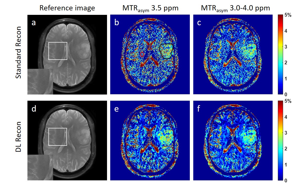
Figure 1. The reference images using standard
recon (a) and DL Recon (d). The MTRasym map at 3.5 ppm (b,e) and
averaged between 3.0-4.0 ppm (c,f) using standard recon (b,c) and DL recon
(e,f) of a glioma patient. The tumor ROI (black) and the background ROI of
homogenous brain tissue (red) was shown in (a) as an example.
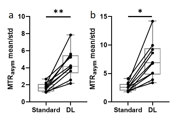
Figure 3. The tumor ROI-averaged MTRasym divided by the standard
deviation of the background ROI at 3.5 ppm (a) and 3.0-4.0 ppm (b) using
standard recon and DL Recon were compared for all patients. (a) p = 0.002. (b)
p = 0.016. **: p < 0.01. *: p < 0.05.