Philip S Boyd1, Johannes Breitling1, Stephanie Laier2, Karin Mueller-Decker2, Andrey Glinka3, Mark E Ladd1, Peter Bachert1, and Steffen Goerke1
1Division of Medical Physics in Radiology, German Cancer Research Center (DKFZ), Heidelberg, Germany, 2Center for Preclinial Research, Core Facility Tumor Models, German Cancer Research Center (DKFZ), Heidelberg, Germany, 3Division of Molecular Embryology, German Cancer Research Center (DKFZ), Heidelberg, Germany
1Division of Medical Physics in Radiology, German Cancer Research Center (DKFZ), Heidelberg, Germany, 2Center for Preclinial Research, Core Facility Tumor Models, German Cancer Research Center (DKFZ), Heidelberg, Germany, 3Division of Molecular Embryology, German Cancer Research Center (DKFZ), Heidelberg, Germany
The
presented method allows calculation of reliable pH maps in the presence of
varying concentration, superimposing CEST signals, magnetization transfer and
spillover dilution. Applicability in vivo was demonstrated in mice showing an
average intracellular pH in tumors of approximately 7.
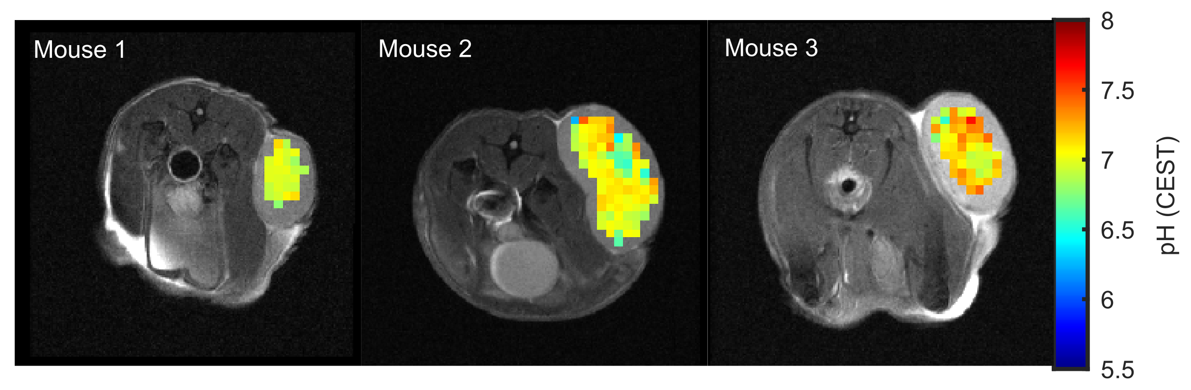
Figure 3:
In vivo application of the calibrated pH-CEST technique in tumor-bearing mice. pH
values of 6.97 ± 0.09, 7.00 ± 0.20 and 7.13 ± 0.19 were found in the subcutaneous lesions
of three DLD xenografted nude mice.
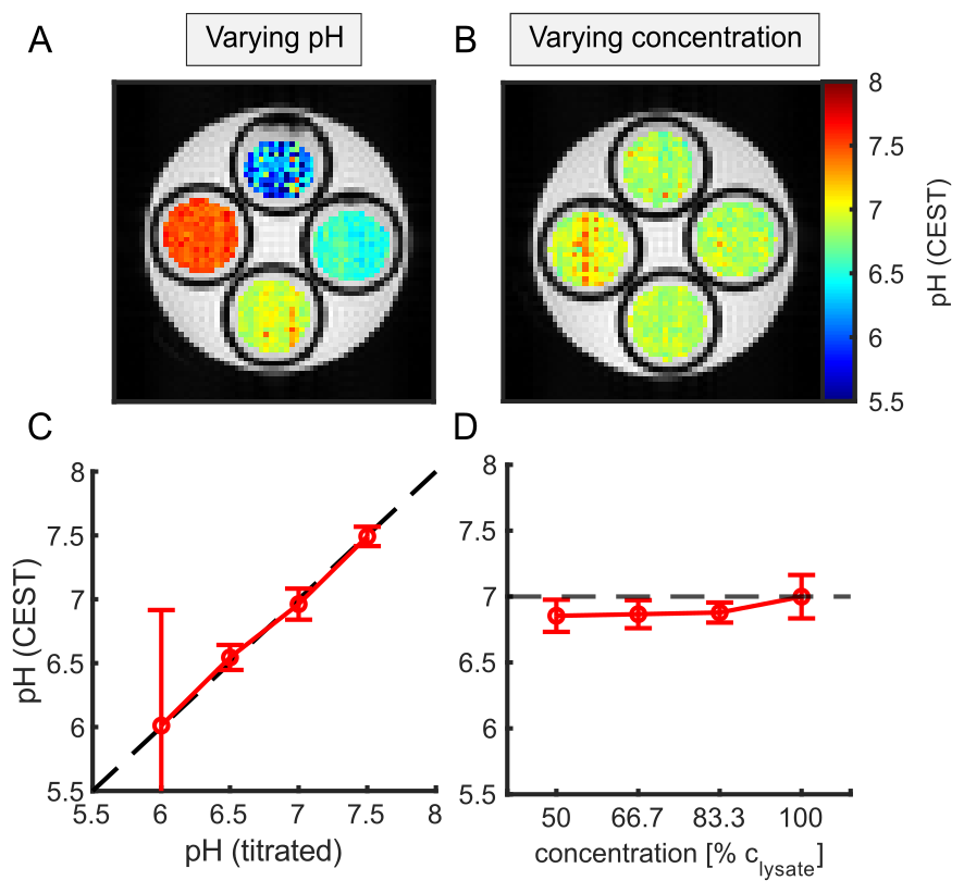
Figure 2:
Correlation of calculated pH from CEST-MRI with titrated pH (A,C) and tissue
concentration (B,D) of porcine brain lysate. C: Correlation of the titrated pH
and the mean pH values calculated from the CESTratio. D: Mean pH
values calculated from the CESTratio as a function of concentration.