Ran Sui1,2,3, Lin Chen1,2, Yuguo Li1,2, Jianpan Huang4, Kannie W.Y Chan2,4, Xiang Xu5, Peter C.M.van Zijl1,2, and Jiadi Xu1,2
1F.M. Kirby Research Center for Functional Brain Imagin, Kennedy Krieger Institute, Baltimore, MD, United States, 2Russell H. Morgan Department of Radiology and Radiological Science, Johns Hopkins University School of Medicine, Baltimore, MD, United States, 3Department of Biomedical Engineering, Johns Hopkins University, Baltimore, MD, United States, 4Department of Biomedical Engineering, City University of Hong Kong, Hong Kong, China, 5BioMedical Engineering and Imaging Institute, Icahn School of Medicine at Mount Sinai, New York, NY, United States
1F.M. Kirby Research Center for Functional Brain Imagin, Kennedy Krieger Institute, Baltimore, MD, United States, 2Russell H. Morgan Department of Radiology and Radiological Science, Johns Hopkins University School of Medicine, Baltimore, MD, United States, 3Department of Biomedical Engineering, Johns Hopkins University, Baltimore, MD, United States, 4Department of Biomedical Engineering, City University of Hong Kong, Hong Kong, China, 5BioMedical Engineering and Imaging Institute, Icahn School of Medicine at Mount Sinai, New York, NY, United States
Acquire whole-brain amide CEST mapping at 3T MRI by using steady-state radial sampling CEST with PROPELLOR-based sampling
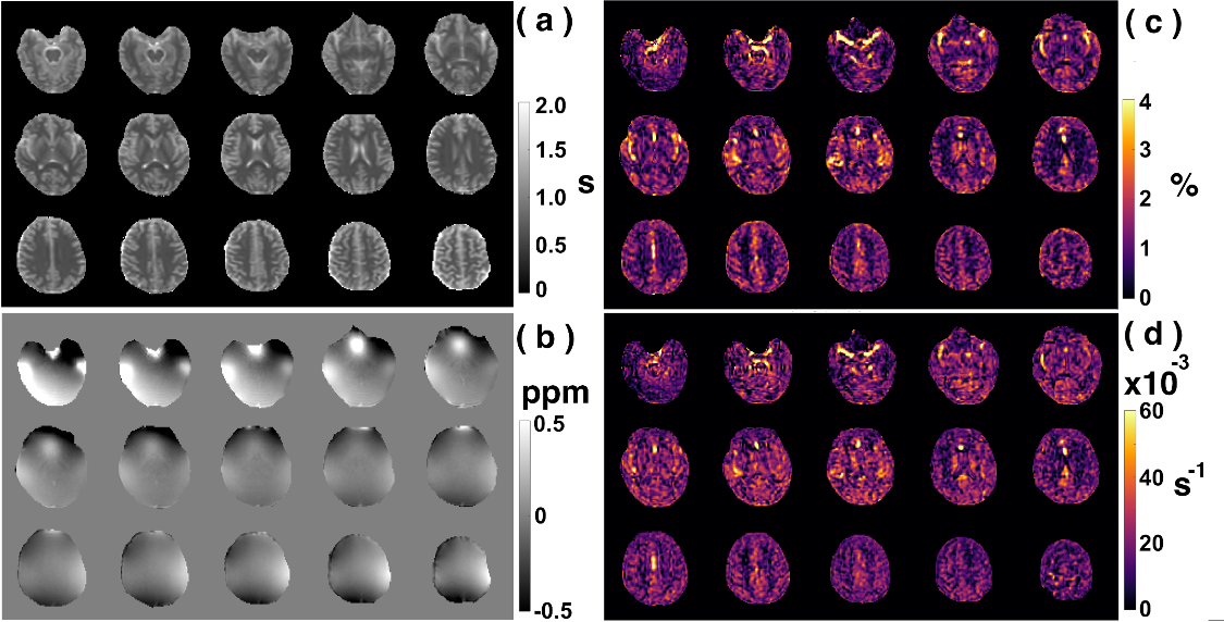
Figure 3: Typical multi-slice human brain images acquired with starCEST at 3T. (a) T1 maps by look-locker sequence; (b) B0 maps acquired with dual-echo sequence, The (c) amideCEST maps (ΔZamide) and the corresponding (d) apparent relaxation rate (Ramide) maps extracted with the PLOF method by including the B0 and T1 maps, respectively.
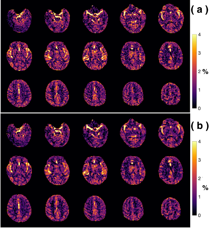
Comparison of the amideCEST maps without and with MLSVD post-processing. (a) The original amide CEST maps extracted using PLOF without MLSVD denoising. (b) MLSVD denoising applied with truncation numbers 48, 48 and 10.