Helen Sporkin1, Toral Patel2, Christopher Schumann2, Christopher Kramer2,3, and Craig Meyer1,3
1Biomedical Engineering, University of Virginia, Charlottesville, VA, United States, 2Cardiovascular Medicine, University of Virginia, Charlottesville, VA, United States, 3Radiology and Medical Imaging, University of Virginia, Charlottesville, VA, United States
1Biomedical Engineering, University of Virginia, Charlottesville, VA, United States, 2Cardiovascular Medicine, University of Virginia, Charlottesville, VA, United States, 3Radiology and Medical Imaging, University of Virginia, Charlottesville, VA, United States
Our preliminary findings in six patients show success in imaging PAD patients with CrCEST and exercise-induced hyperemia ASL in the same protocol. Further recruitment is necessary to establish findings linking perfusion, metabolism, and function.
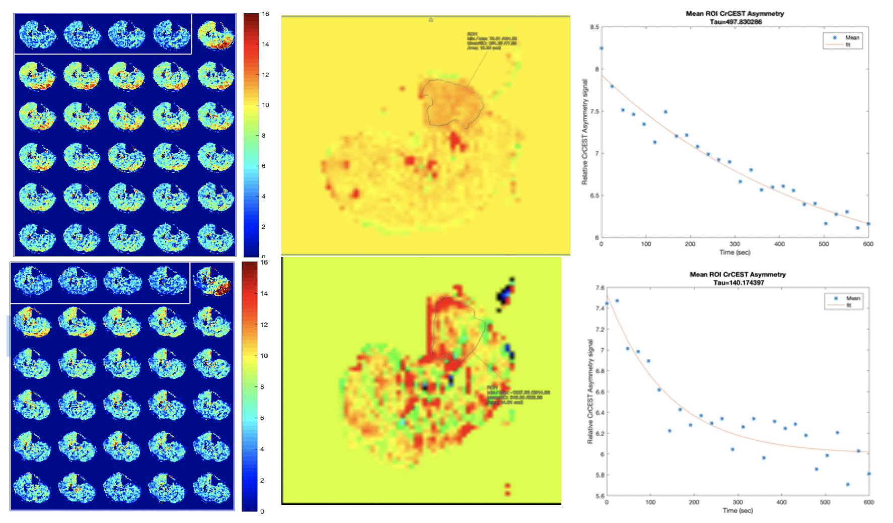
Figure 1: (Top row) CrCEST time course imaging (left) ,and ASL (middle), and overall CrCEST decay (right) in a patient with an ABI of 0.68 prior to endovascular revascularization.(Bottom row) CrCEST time course imaging (left) , ASL (middle), and overall CrCEST decay (right) CrCEST time course imaging and ASL in the same patient 6 months after revascularization, showing changes in both metabolism and perfusion.
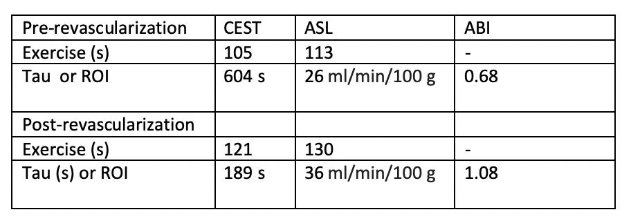
Table 1: CEST and ASL evaluation pre and post revascularization in patient 1. ASL and CEST measurements are reported as a mean of the muscle ROI in which the greatest perfusion was seen in the ASL images.