Jeehun Kim1,2, Chaoyi Zhang3, Mingrui Yang1, Hongyu Li3, Mei Li1, Richard Lartey1, Leslie Ying3,4, and Xiaojuan Li1
1Department of Biomedical Engineering, Program of Advanced Musculoskeletal Imaging (PAMI), Cleveland Clinic, Cleveland, OH, United States, 2Department of Electrical Engineering, Case Western Reserve University, Cleveland, OH, United States, 3Electrical Engineering, University at Buffalo, State University of New York, Buffalo, NY, United States, 4Biomedical Engineering, University at Buffalo, State University of New York, Buffalo, NY, United States
1Department of Biomedical Engineering, Program of Advanced Musculoskeletal Imaging (PAMI), Cleveland Clinic, Cleveland, OH, United States, 2Department of Electrical Engineering, Case Western Reserve University, Cleveland, OH, United States, 3Electrical Engineering, University at Buffalo, State University of New York, Buffalo, NY, United States, 4Biomedical Engineering, University at Buffalo, State University of New York, Buffalo, NY, United States
Compared to reference images, cartilage T1ρ
coefficients of variation < 3.5% was achieved with prospective acceleration
factor of 8, which reduced the scan time to 3 minutes, for subjects with and
without osteoarthritis.
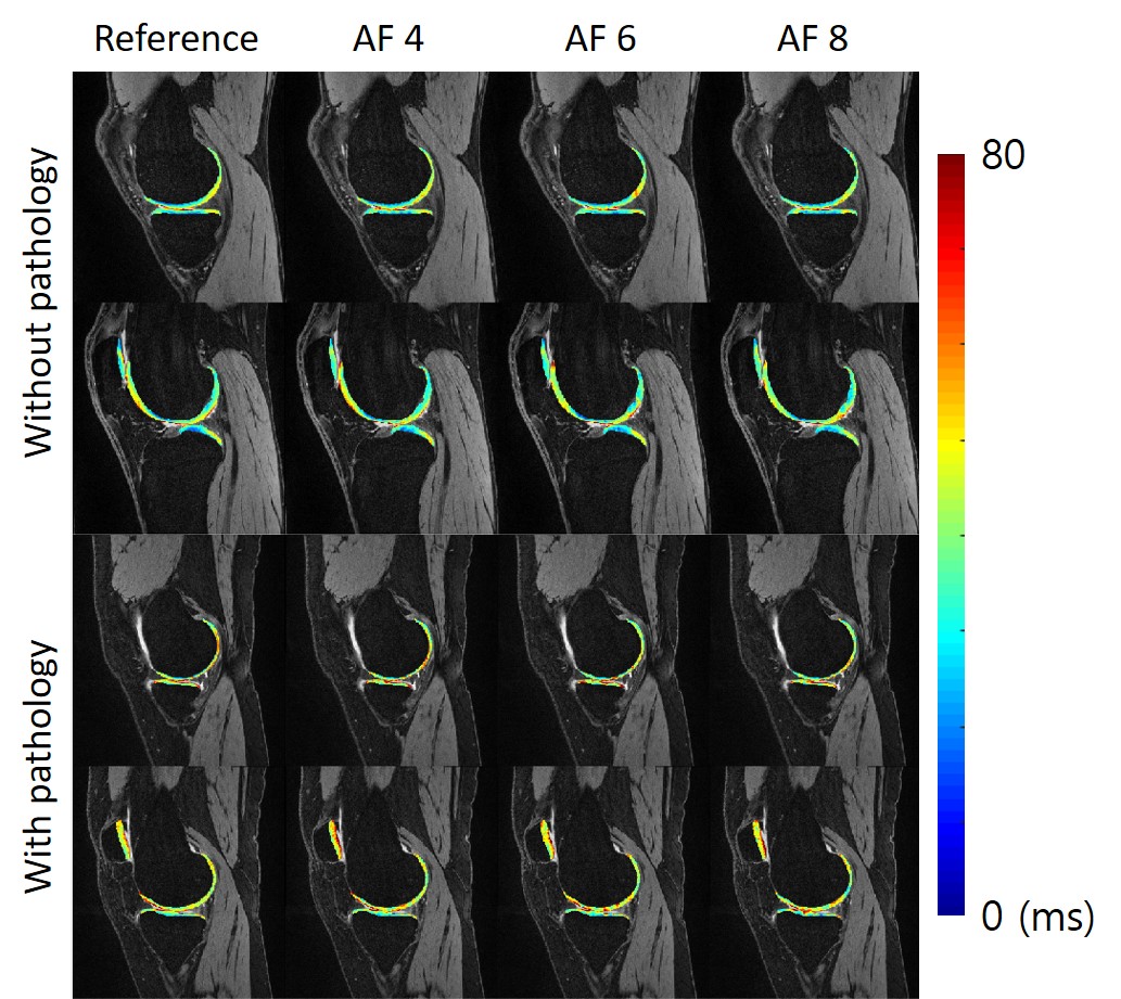
Figure 4 Sample images of reference and
accelerated T1ρ map. The T1ρ map
of cartilage compartments was overlaid to the corresponding DESS images for
better visualization.
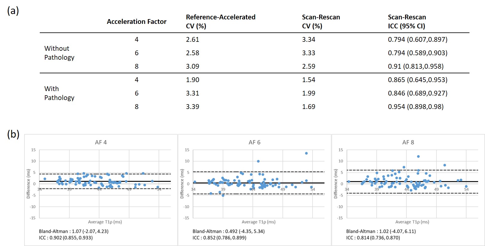
Figure 3 (a) shows the CV between reference
and accelerated T1ρ value, scan-rescan CV, and
scan-rescan ICC with a 95% confidence interval. (b) shows the Bland-Altman plot
between accelerated and reference T1ρ value. Each entry corresponds
to an average value of a cartilage compartment in a subject. ICC was calculated for
each AF.