Gabriele Bonanno1,2,3, Tom Hilbert4,5,6, Patrick Leibig7, and Tobias Kober4,5,6
1Advanced Clinical Imaging Technology,Siemens Healthcare AG, Bern, Switzerland, 2Translational Imaging Center, sitem-insel AG, Bern, Switzerland, 3Departments of Radiology and Biomedical Research, University of Bern, Bern, Switzerland, 4Advanced Clinical Imaging Technology, Siemens Healthcare AG, Lausanne, Switzerland, 5Department of Radiology, University Hospital (CHUV) and University of Lausanne (UNIL), Lausanne, Switzerland, 6LTS5, École Polytechnique Fédérale de Lausanne, Lausanne, Switzerland, 7Siemens Healthcare GmbH, Erlangen, Germany
1Advanced Clinical Imaging Technology,Siemens Healthcare AG, Bern, Switzerland, 2Translational Imaging Center, sitem-insel AG, Bern, Switzerland, 3Departments of Radiology and Biomedical Research, University of Bern, Bern, Switzerland, 4Advanced Clinical Imaging Technology, Siemens Healthcare AG, Lausanne, Switzerland, 5Department of Radiology, University Hospital (CHUV) and University of Lausanne (UNIL), Lausanne, Switzerland, 6LTS5, École Polytechnique Fédérale de Lausanne, Lausanne, Switzerland, 7Siemens Healthcare GmbH, Erlangen, Germany
High isotropic resolution and full-brain T1ρ maps obtained at the scanner from
an accelerated and optimized FLASH sequence are demonstrated to provide
high repeatability and reproducibility in a healthy volunteer cohort.
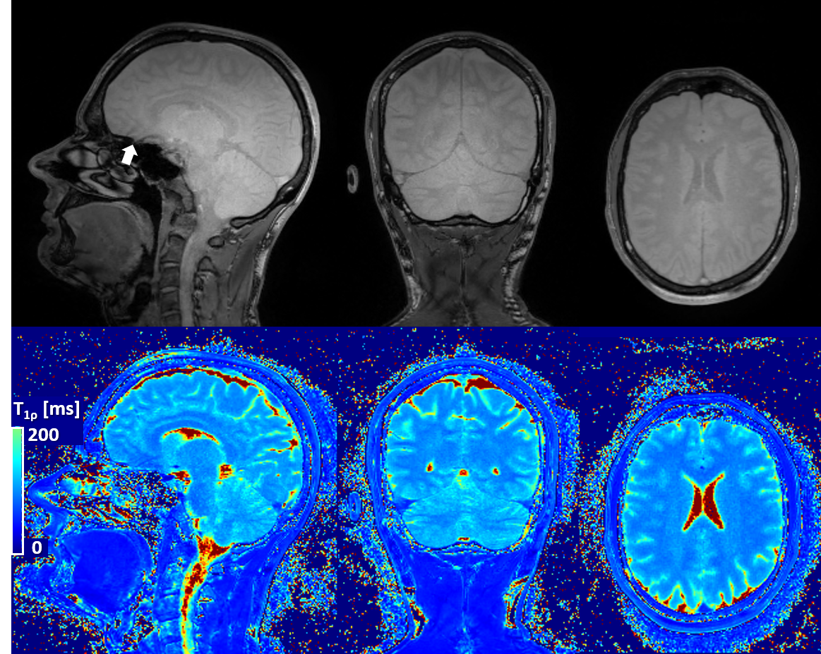
Figure 3
Representative T1ρ-prepared images (only SL-time=30 ms shown in top row) and T1ρ map (bottom row) obtained from the same acquisition. Homogenous T1ρ contrast can be observed in the whole brain, even in lower structures for SL-time=30 ms. Occasionally some artifacts may be observed above the nasal cavity due to air-tissue boundaries (arrow). Quantification of T1ρ also shows good contrast and homogeneity throughout the brain.
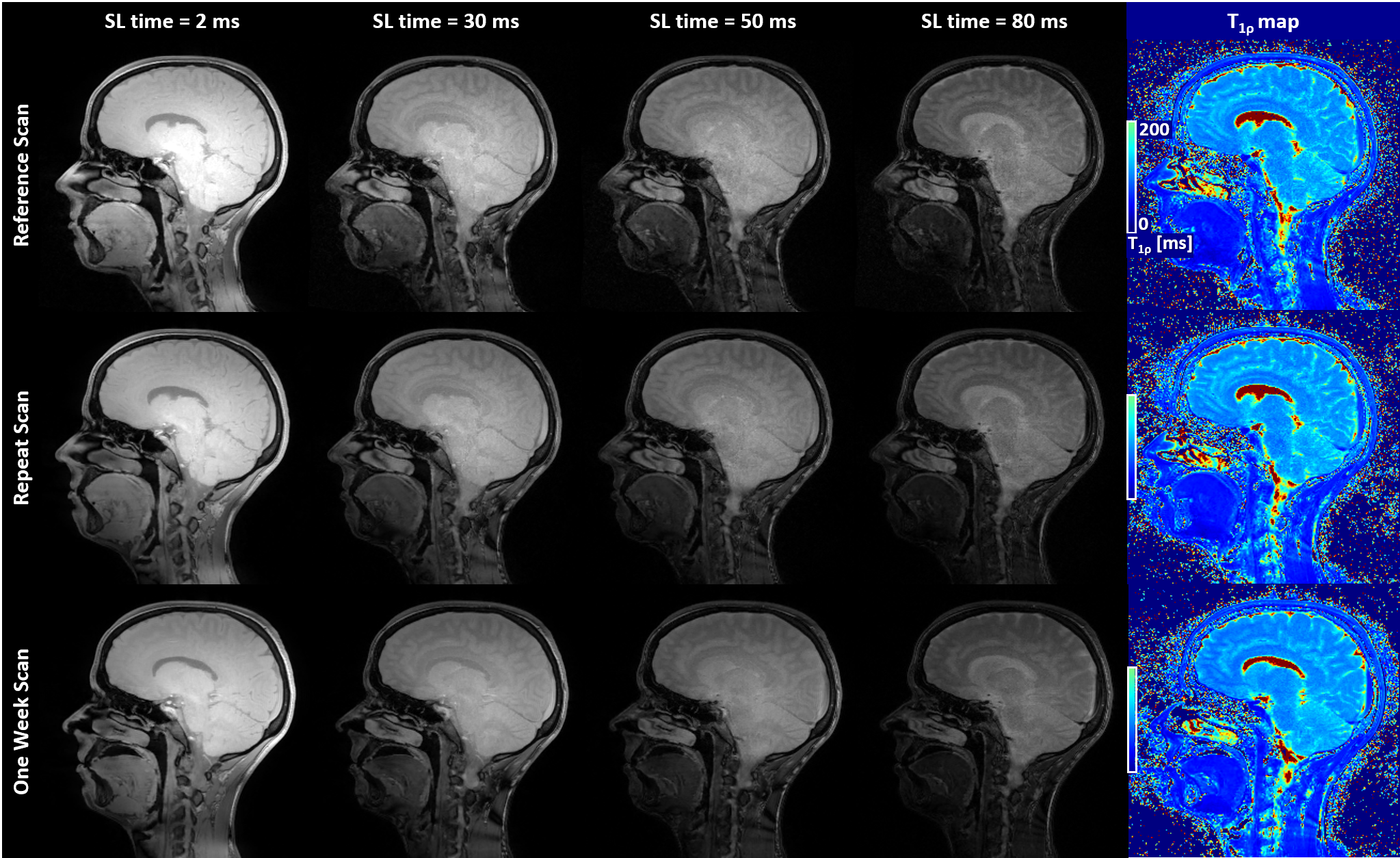
Figure 4
Example sagittal slices from T1ρ-prepared volumes and the resulting map in the same subject during Reference, Repeat and One-Week scan show increased T1ρ weighting as function of SL-time as well as good visual repeatability and reproducibility of image quality between scans.