Gopal Varma1, Aaron K Grant1, Olivier M Girard2, Guillaume Duhamel2, and David C Alsop1
1Radiology, Division of MR Research, Beth Israel Deaconess Medical Center, Harvard Medical School, Boston, MA, United States, 2CNRS, CRMBM, Aix-Marseille Univ, Marseille, France
1Radiology, Division of MR Research, Beth Israel Deaconess Medical Center, Harvard Medical School, Boston, MA, United States, 2CNRS, CRMBM, Aix-Marseille Univ, Marseille, France
Quantitative inhomogeneous Magnetization Transfer of brain and spinal cord tissues showed differences in free pool longitudinal relaxation rate R1a, bound pool transverse relaxation time T2b, and dipolar relaxation time T1d between samples from donors with and without multiple sclerosis.
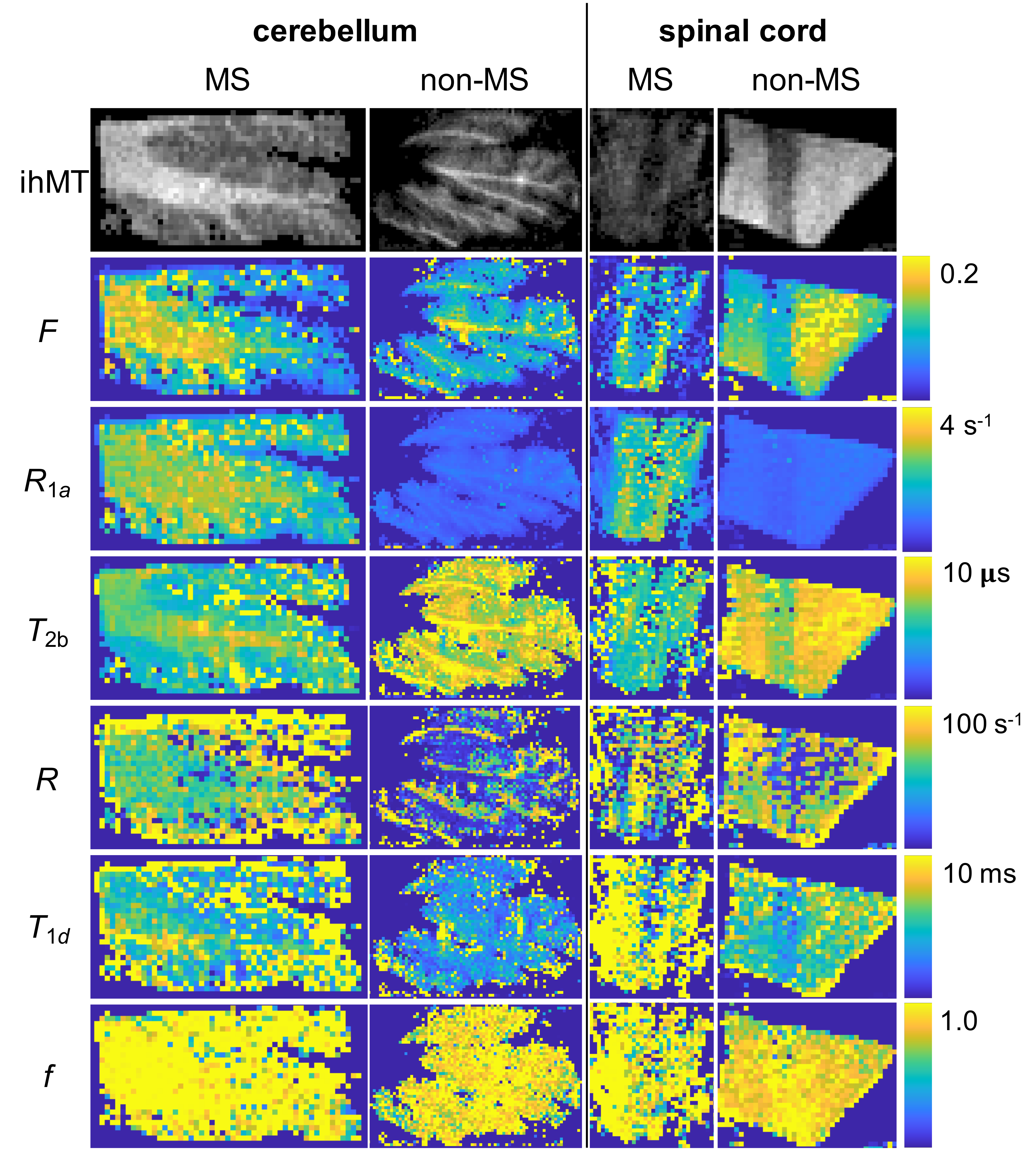
Figure 4 IhMT images and maps of selected qihMT parameters output from global fits to ihMT data acquired with optimized protocol in central nervous system tissue from donors with and without MS. The ihMT images in the top row are formed from the difference between the summation of data prepared with single or dual frequency RF irradiation applied off-resonance. Color bars on the right relate to the maps of qihMT parameters in the same row all with lowest levels of 0 and highest indicated. All images and maps were resized to twice their resolution by nearest-neighbor interpolation.
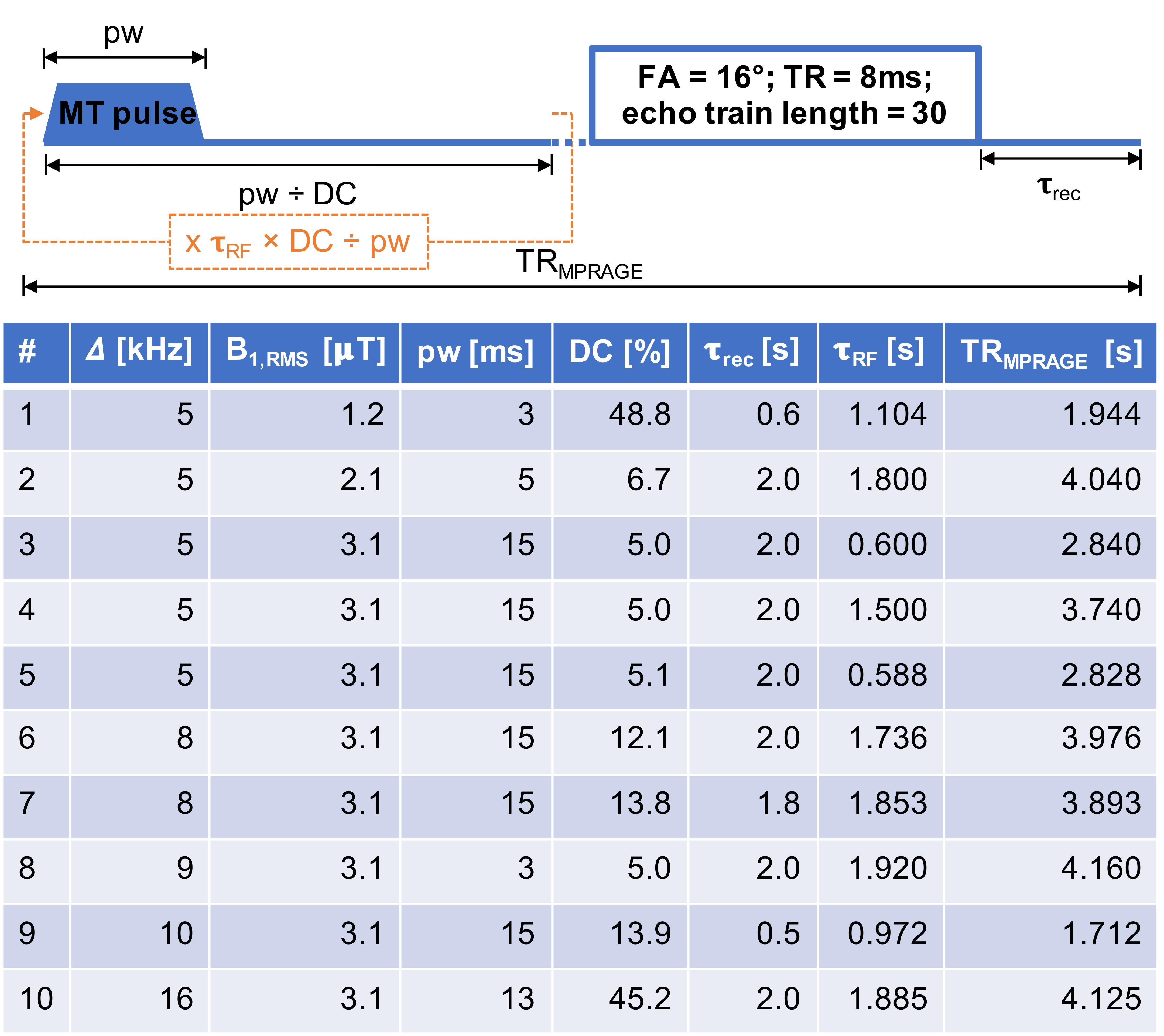
Figure 1 Schematic of MT sequence (top) for ihMT experiments illustrating some of the scan parameters open to optimization and table of optimal protocol of scan parameters (bottom) from CRLB analysis. Optimized parameters included: MT preparation duration (𝛕RF), recovery time (𝛕rec), root mean square B1 (B1,RMS) over the MT preparation, and frequency (𝛥), pulse width (pw), and duty cycle (DC) of MT pulses.
