Anliang Chen1, Ailian Liu1, Jiazheng Wang2, Zhiwei Shen2, Deshuo Dong1, Wan Dong1, Yuhui Liu1, Qingwei Song1, and Renwang Pu1
1Radiology, The First Affiliated Hospital of Dalian Medical University, Dalian, China, 2Philips Healthcare, Beijing, China
1Radiology, The First Affiliated Hospital of Dalian Medical University, Dalian, China, 2Philips Healthcare, Beijing, China
APTw imaging combined with T1 map can effectively
reflect lesion changes between rectal
cancer with and without lymph node metastasis.
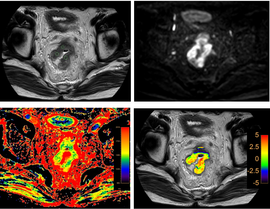
Figure 1. A 65-year-old
male patient with lymph node metastasis of rectal cancer. T2W image (a), DWI (b), T1 map (c), and APTw-T2W
fusion image (d) to show the lesion of rectal cancer with lymph
node metastasis. Three ROI of
rectal cancer were showed on T2W image. APT and T1 values of the ROI were 0.89%,
0.45%, 0.97% and 1579.90ms, 1449.80ms, 1496.05ms.
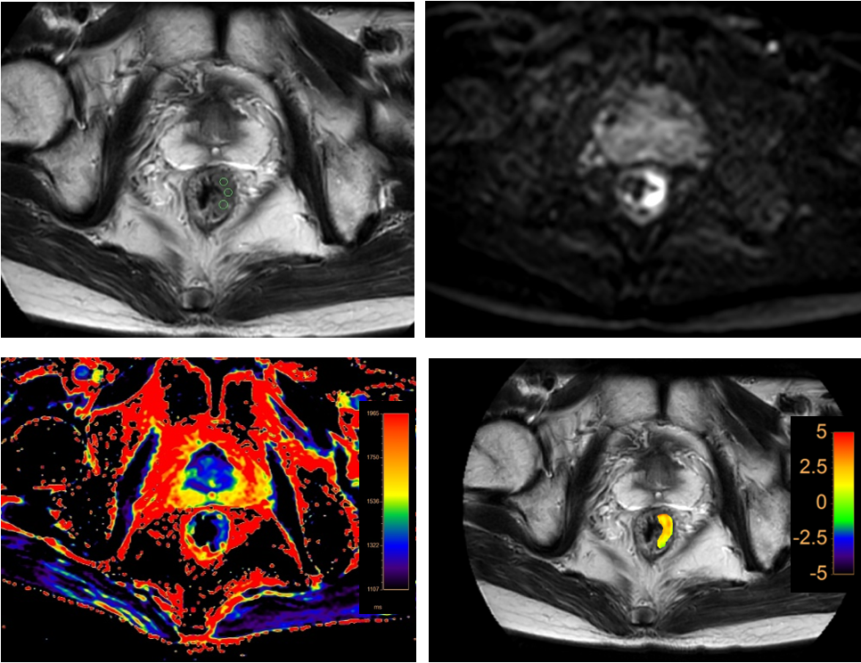
Figure 2. A 64-year-old male patient without lymph
node metastasis of
rectal cancer. T2W image (a), DWI (b), T1 map (c), and APTw-T2W fusion image (d) to
show the lesion of rectal cancer without lymph node metastasis. Three ROI of rectal
cancer were showed on T2W image. APT and T1 values of the ROI were 2.03%, 2.64%,
1.73% and 1371.23ms, 1337.87ms, 1353.56ms.