Ying-Hua Chu1, Yi-Cheng Hsu1, and Patrick Alexander Liebig2
1MR Collaboration, Siemens Healthcare Ltd., Shanghai, China, 2Siemens Healthcare GmbH, Erlangen, Germany
1MR Collaboration, Siemens Healthcare Ltd., Shanghai, China, 2Siemens Healthcare GmbH, Erlangen, Germany
We
proposed a new B0/B1 correction method that undersamples the number of offsets. For APTw imaging in brain, the method halved the error caused by B1 inhomogeneity at 3T with 157% scan time.
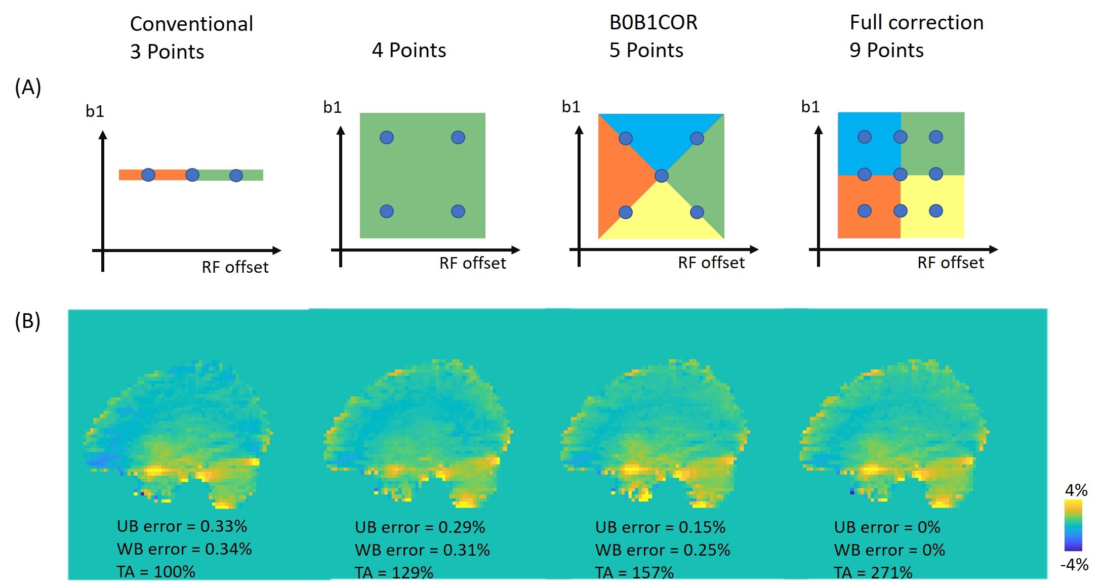
Figure2: (A)
The illustration of B1 and RF offsets sampling and correction methods for
conventional, 4 points, B0B1CAR, and full correction methods, respective. (B)
The sagittal APTw images with B0 and B1 correction by the corresponding
methods. The mean errors in the upper brain region (UB error) and the whole
brain region (WB error) are measured by the mean difference[LP(DMRS1] with the full correction method. The relative total acquisition times
(TA) are compared with the conventional method.
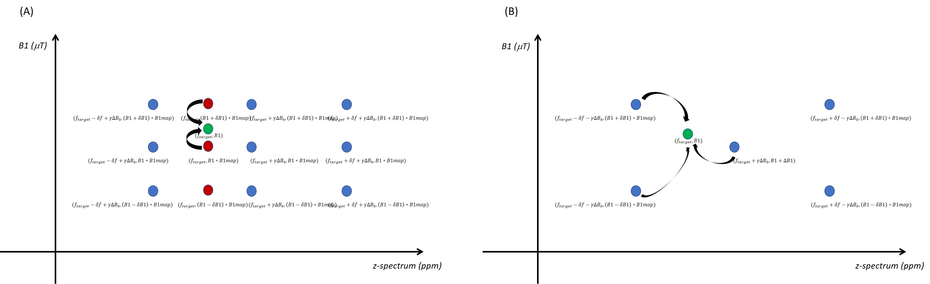
Figure1: (A) Conventional method for CEST B0 and B1 correction. First, the data at the
targeted z-spectrum frequency was interpolated at each B1 value. Then, the data
at the targeted frequency and B1 were interpolated from the B0 corrected data.
(B) B0B1COR method for CEST B0 and B1 correction. The data at the targeted
z-spectrum frequency and B1 was interpolated directly from nearby sampled
z-spectrum at different B1 values.