Zhuonan Wang1, Victoria J Williams2, Kimberly A Stephens3, Chan-Mi Kim3, Ming Zhang4, and David Salat3
1PET/CT Unit, Medical Imaging, The First Affiliated Hospital of Xi'an Jiaotong University, Xi'an, China, 2Alzheimer's Clinical and Translational Research Unit, Department of Neurology, Massachusetts General Hospital, Charlestown, MA, United States, 3Athinoula A. Martinos Center for Biomedical Imaging, Department of Radiology, Massachusetts General Hospital, Charlestown, MA, United States, 4Medical Imaging, The First Affiliated Hospital of Xi'an Jiaotong University, Xi'an, China
1PET/CT Unit, Medical Imaging, The First Affiliated Hospital of Xi'an Jiaotong University, Xi'an, China, 2Alzheimer's Clinical and Translational Research Unit, Department of Neurology, Massachusetts General Hospital, Charlestown, MA, United States, 3Athinoula A. Martinos Center for Biomedical Imaging, Department of Radiology, Massachusetts General Hospital, Charlestown, MA, United States, 4Medical Imaging, The First Affiliated Hospital of Xi'an Jiaotong University, Xi'an, China
Reduced DMN functional connectivity in
those with MCI compared to cognitively healthy controls, the extent to which
was differentially regionally related to white matter lesion volume and may indicate
a vascular etiology to subtle impairment in MCI.
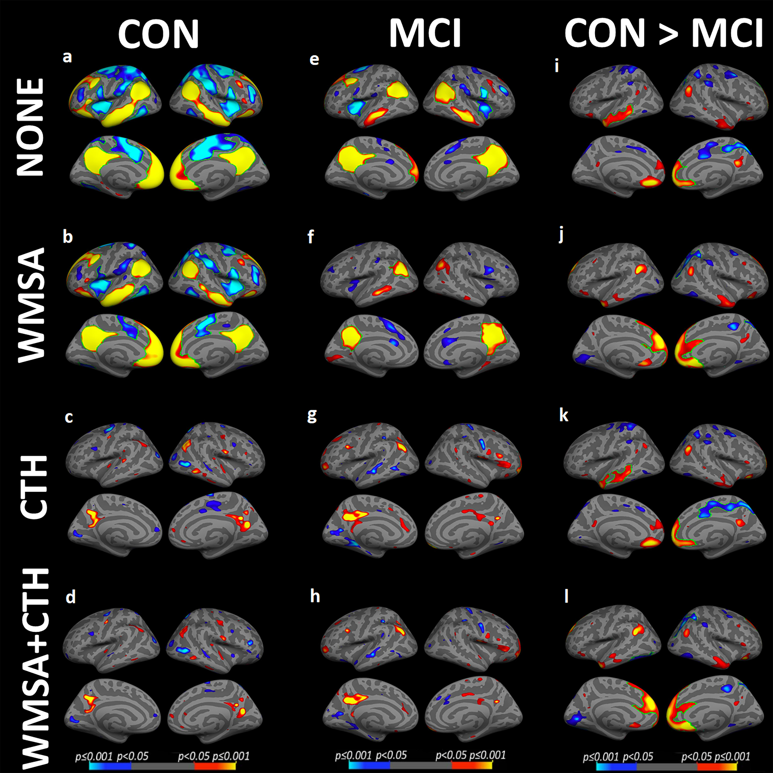
Functional connectivity maps are shown for (1) cognitively healthy controls (CON), (2) Mild cognitive impairment (MCI) group and groups comparison : (a,e,i) without regressors, (b,f,j) regressing out white matter signal abnormalities (WMSA), (c,g,k) regressing out cortical thickness (CTH), and (d,h,l) regressing out both. Warm colors indicate significant regional positive correlations with the precuneus-seed (a-h) and stronger DMN connectivity in CON, and cool colors indicate regions of anticorrelation (a-h) and stronger DMN connectivity in MCI.