Vadim Zotev1, Aki Tsuchiyagaito1, and Jerzy Bodurka1,2
1Laureate Institute for Brain Research, Tulsa, OK, United States, 2Stephenson School of Biomedical Engineering, University of Oklahoma, Norman, OK, United States
1Laureate Institute for Brain Research, Tulsa, OK, United States, 2Stephenson School of Biomedical Engineering, University of Oklahoma, Norman, OK, United States
Our study provides evidence using simultaneous EEG-fMRI that EEG neurofeedback training targeting frontal alpha EEG asymmetry can engage and influence the emotional brain circuitry in patients with major depressive disorder.
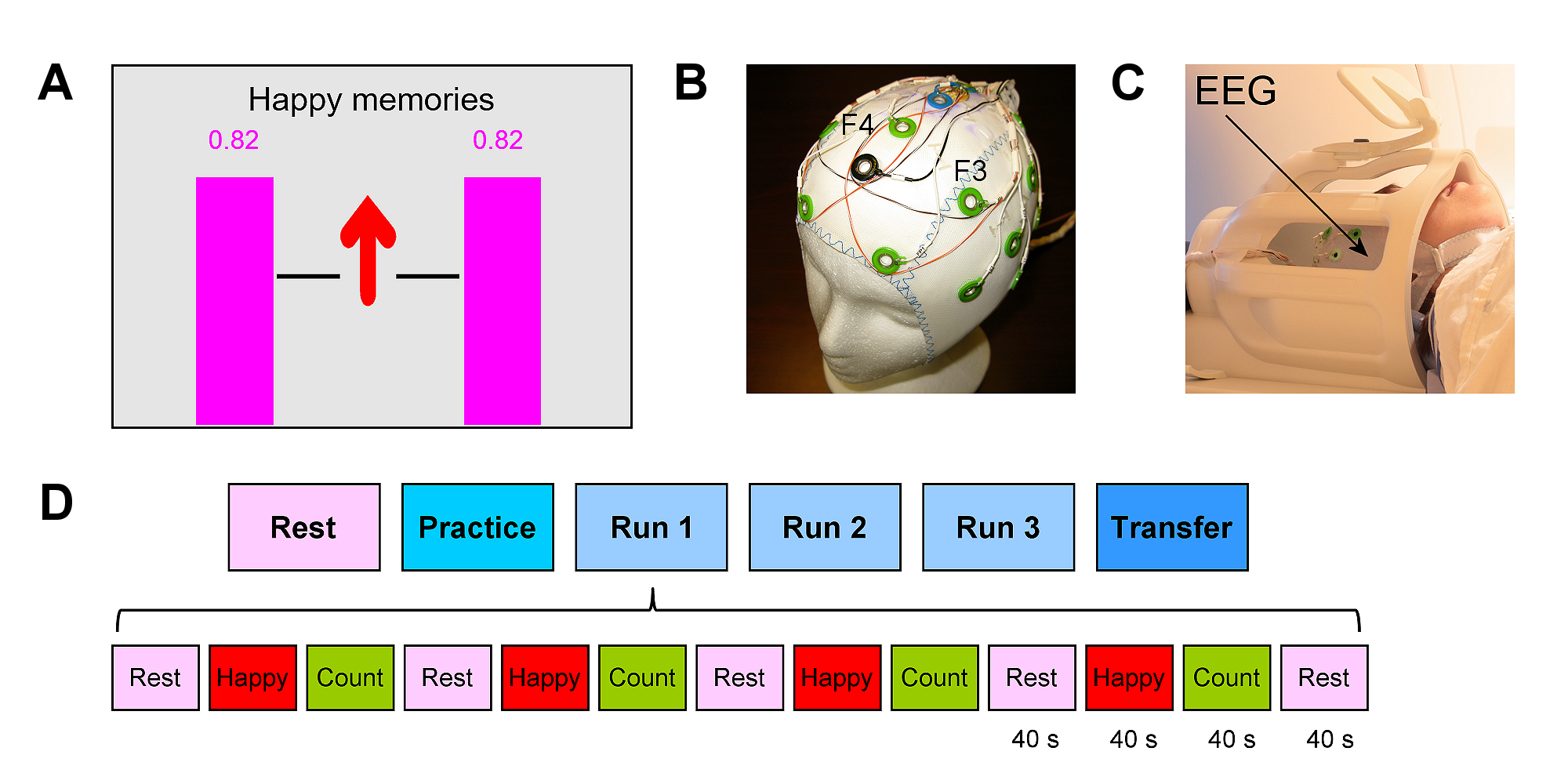
Figure 1. A) EEG neurofeedback GUI screen with variable-height
magenta neurofeedback bars. B) Modified MR-compatible EEG cap for improved
EEG-nf during fMRI. Frontal channels F3 and F4 are used to provide FAA-based EEG-nf.
C) Simultaneous EEG-fMRI. D) Experimental protocol with six EEG-fMRI runs,
abbreviated as RE, PR, R1, R2, R3, and TR. The five task runs consist of 40-s-long
blocks of Rest, Happy Memories, and Count conditions, abbreviated as R, H, and
C.
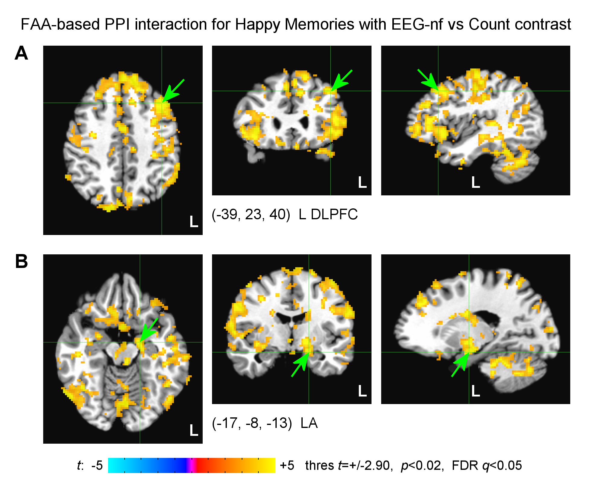
Figure
4.
Statistical maps of the FAA-based PPI interaction effect for the Happy Memories
with EEG-nf vs Count condition contrast for the EG. The individual results were
averaged across the four EEG-nf runs. The maps are shown in the Talairach
space. A positive effect means a stronger temporal correlation between the FAA
time course and BOLD activity during the EEG-nf task compared to the control
task. A) Left dorsolateral prefrontal cortex (L DLPFC) region. B) Left amygdala
(LA) region.