Shin-Eui Park1, Gwang-Won Kim2, Yun-Hyeon Kim3, and Gwang-Woo Jeong3
11Advanced Institute of Aging Science, Chonnam National University, Gwangju, Korea, Republic of, 2Department of Psychiatry, Massachusetts General Hospital and Harvard Medical School, Boston, MA, United States, 3Department of Radiology, Chonnam National University Medical School, Gwangju, Korea, Republic of
11Advanced Institute of Aging Science, Chonnam National University, Gwangju, Korea, Republic of, 2Department of Psychiatry, Massachusetts General Hospital and Harvard Medical School, Boston, MA, United States, 3Department of Radiology, Chonnam National University Medical School, Gwangju, Korea, Republic of
Our findings indicate abnormal connection with superior temporal cortex and inferior parietal cortex caused by obsessive-compulsive symptom.
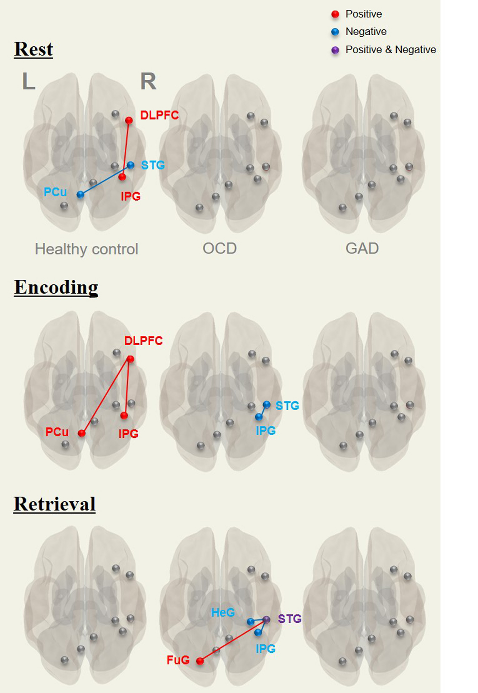
Different brain functional connectivity patterns in patients with OCD and
GAD and in healthy control participants during memory tasks. Red lines
represent positive connectivity and blue lines indicate negative connectivity. None
of the functional connectivity patterns were observed in patients with GAD. Details
are shown in Table 4. DLPFC, dorsolateral prefrontal cortex; STG, superior
temporal gyrus; IPG, inferior parietal gyrus; PCu, precuneus; HeG, Heschl
gyrus; FuG, fusiform gyrus. L=left; R=right.
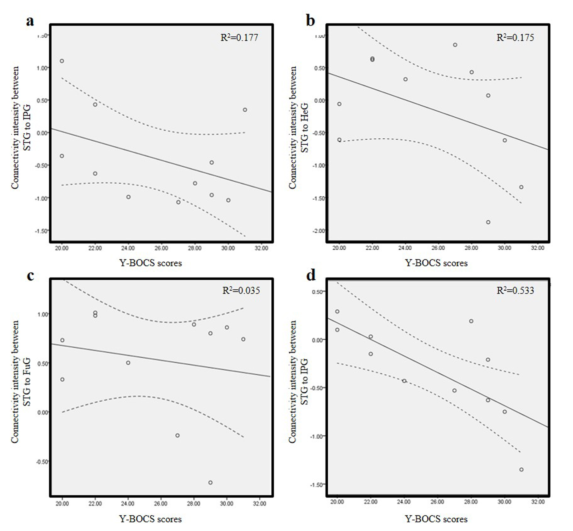
Correlations between Y-BOCS scores and functional connectivity(FC) during explicit, verbal memory tasks: in encoding period, STG-IPG(r=-0.421,p=0.011)(a); in retrieval period, STG-HeG(r=-0.418,p=0.200)(b), STG-FuG(r=-0.189,p=0.581)(c), and STG-IPG(r=-0.421,p=0.011)(d). Note
that only FC of STG-IPG was negatively correlated with
Y-BOCS scores. Curved dotted line bands indicate 95% confidence intervals.
Y-BOCS, Yale-Brown Obsessive Compulsive Scale; STG,
superior temporal gyrus; IPG, inferior parietal gyrus; HeG, Heschl gyrus; FuG,
fusiform gyrus.