Karthik R Sreenivasan1, Xiaowei Zhuang1, Zhengshi Yang1, Dietmar Cordes1, Aaron Ritter1, Jessica Caldwell1, Zoltan Mari1, and Virendra Mishra1
1Cleveland Clinic Lou Ruvo Center for Brain Health, Las Vegas, NV, United States
1Cleveland Clinic Lou Ruvo Center for Brain Health, Las Vegas, NV, United States
The results of our study demonstrate that despite not observing overall
global or local network differences Parkinson’s disease patients with and without
freezing of gait exhibit a clear shift in the topological organization when
compared to the controls.
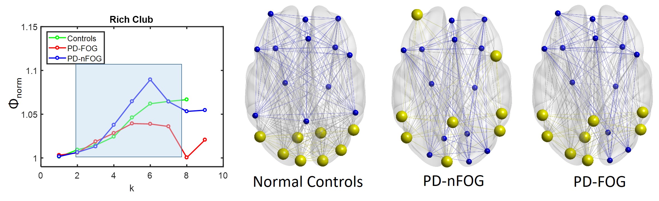
Figure 3. Rich-clubs are reorganized in the PD groups when compared to
controls. (A) The Rich-club regime for NC, PD-nFOG, and PD-FOG are shown in the blue-shaded
box. (B) Rich-club nodes are indicated in yellow color and non-rich-club nodes
are shown in blue color for NC, PD-nFOG, and PD-FOG. Red circle – default mode
network; purple circle – frontoparietal network; green circle – visual network.
‘k’ is the nodal degree. Visualized using BrainNet Viewer10
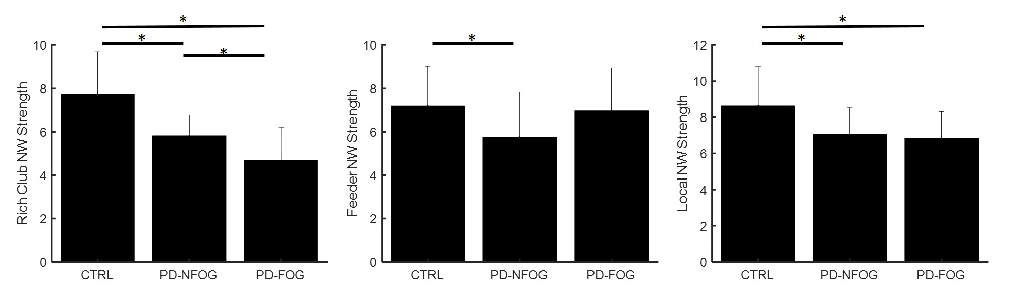
Figure 4. The difference in rich-club connectivity. Rich-club network strength,
feeder network strength, and local network strength are plotted as bar plots
for NC, PD-nFOG, and PD-FOG groups. Rich-club network – edges between rich-club
nodes; feeder network – edges connecting non-rich-club nodes and rich club
nodes; local network – edges between non-rich-club nodes. * indicates
statistical significance (pcorr<0.05)