Li Jiang1,2, Stephanie Chen3, Lorenna Vidal4, Jiachen Zhuo1,2, Rao Gullapalli1,2, and Prashant Raghavan2
1Center for Advanced Imaging Research, University of Maryland Baltimore, Baltimore, MD, United States, 2Department of Diagnostic Radiology & Nuclear Medicine, University of Maryland Baltimore, Baltimore, MD, United States, 3Department of Neurology, University of Maryland Baltimore, Baltimore, MD, United States, 4Department of Radiology, Children's Hospital of Philadelphia, Philadelphia, PA, United States
1Center for Advanced Imaging Research, University of Maryland Baltimore, Baltimore, MD, United States, 2Department of Diagnostic Radiology & Nuclear Medicine, University of Maryland Baltimore, Baltimore, MD, United States, 3Department of Neurology, University of Maryland Baltimore, Baltimore, MD, United States, 4Department of Radiology, Children's Hospital of Philadelphia, Philadelphia, PA, United States
TLE patients showed decreased mALFF in right STG and right AG compared to HCs.
Left TLE patients showed a greater decrease in mALFF in the left AG compared to right TLE patients.
Significant positive correlation between averaged mALFF in right STG and clinical comprehension language score.
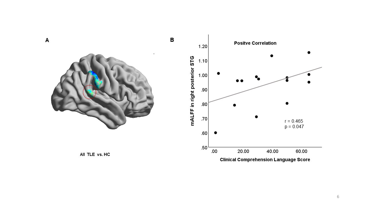
The Pearson
correlation between the language test scores and the mALFF in
the sub-regions within the language eloquent areas. (A) Significant clusters by
comparing all TLE patients and healthy controls. The regions of right superior
temporal gyrus (pSTG) was circled out. (B) Correlation
between the composite percentile scores and mALFF in the
right pSTG.
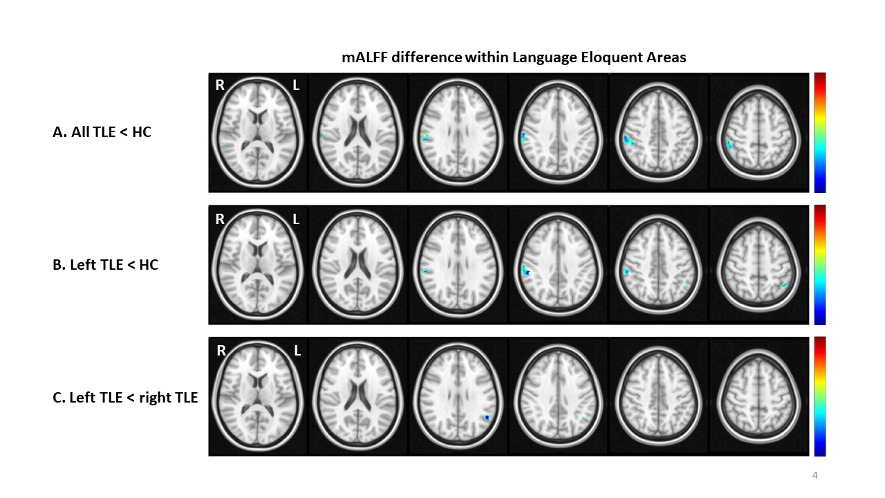
Figure
1: The mALFF difference within language eloquent areas among
groups (A) all TLE
patients and HCs. (B) left TLE and HCs; (C) left TLE
and right TLE patients. The significant difference was defined as
voxel-wise uncorrected p < 0.008, cluster-wise corrected p < 0.05 and
cluster-size > 73 voxels. L:
left hemisphere; R: right hemisphere. Hot color refers to increased mALFF and
cold color refers to decreased mALFF.