Sicong Tu1, Marion Sourty2, Fernando Calamante1, Manojkumar Saranathan3, Ricarda Menke4, Kevin Talbot4, Matthew Kiernan1, and Martin Turner4
1The University of Sydney, Sydney, Australia, 2Université de Strasbourg, Strasbourg, France, 3University of Arizona, Tucson, AZ, United States, 4University of Oxford, Oxford, United Kingdom
1The University of Sydney, Sydney, Australia, 2Université de Strasbourg, Strasbourg, France, 3University of Arizona, Tucson, AZ, United States, 4University of Oxford, Oxford, United Kingdom
In ALS, thalamic
volume is reduced in medial, ventral, and posterior regions, while connectivity
was altered in the anterior region. Connectivity of the mediodorsal nuclei correlated
with disease duration and progression rate, and ventral lateral nuclei with
upper motor neuron dysfunction.
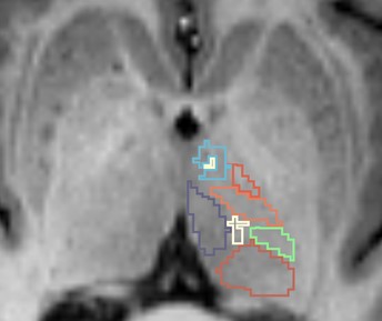
Figure 1. Representative thalamic nuclei segmentation using the
THOMAS pipeline on an individual T1 image.
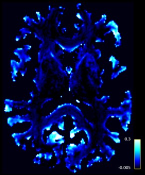
Figure 2. Representative
track-weighted static functional connectivity map for an individual
participant.