Ji Won Bang1, Carlos Parra1, Gadi Wollstein1, Joel S Schuman1, and Kevin C Chan1,2
1Department of Ophthalmology, New York University Grossman School of Medicine, New York, NY, United States, 2Department of Radiology, New York University Grossman School of Medicine, New York, NY, United States
1Department of Ophthalmology, New York University Grossman School of Medicine, New York, NY, United States, 2Department of Radiology, New York University Grossman School of Medicine, New York, NY, United States
Glaucoma patients exhibited altered functional connectivity between the subcortical sleep-inducing hub, the subcortical arousal system, and the occipital cortex. The occipital cortex also showed reduced GABA, suggestive of impaired inhibitory function.
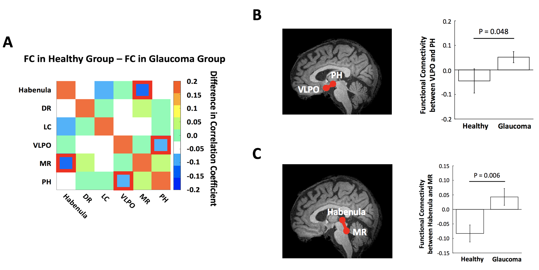
Figure 2. (A) Heatmap showing differences in functional connectivity (FC) (healthy – glaucoma). Red boxes indicate changes in FC that reach significance (P<0.05). FC of VLPO and PH and that of habenula and MR are greater in glaucoma patients than healthy controls. (B) FC between VLPO and PH in healthy vs. glaucoma. (C) FC between habenula and MR in healthy vs. glaucoma. Error bars indicate SEM. (Healthy group: N=22; Glaucoma group: N=38)
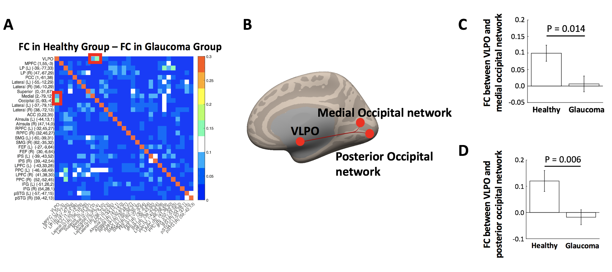
Figure 3. (A) Heatmap showing differences in functional connectivity (FC) (healthy – glaucoma). Red boxes indicate changes in FC that reach significance (P<0.05). FC between VLPO and medial or posterior occipital network is greater in healthy controls than glaucoma patients. (B) Sagittal view showing VLPO, medial and posterior occipital networks. (C) FC between VLPO and medial occipital network in healthy vs. glaucoma. (D) FC between VLPO and posterior occipital network in healthy vs. glaucoma. Error bars indicate SEM. (Healthy group: N=22; Glaucoma group: N=38)