Zhengshi Yang1,2, Cieri Filippo1, Xiaowei Zhuang1,2, Marwan Sabbagh1, Jefferson W. Kinney2, Jeffrey L. Cummings2, Dietmar Cordes1,2,3, and Jessica Z.K. Caldwell1
1Cleveland Clinic Lou Ruvo Center for Brain Health, Las Vegas, NV, United States, 2University of Nevada Las Vegas, Las Vegas, NV, United States, 3University of Colorado Boulder, Boulder, CO, United States
1Cleveland Clinic Lou Ruvo Center for Brain Health, Las Vegas, NV, United States, 2University of Nevada Las Vegas, Las Vegas, NV, United States, 3University of Colorado Boulder, Boulder, CO, United States
Opposite network topological changes were observed from cognitively normal to MCI, and more rapid progression occurred in women than men from MCI to AD. The occipital lobe contributed more in men but frontal lobe contributed more in women in disease progression.
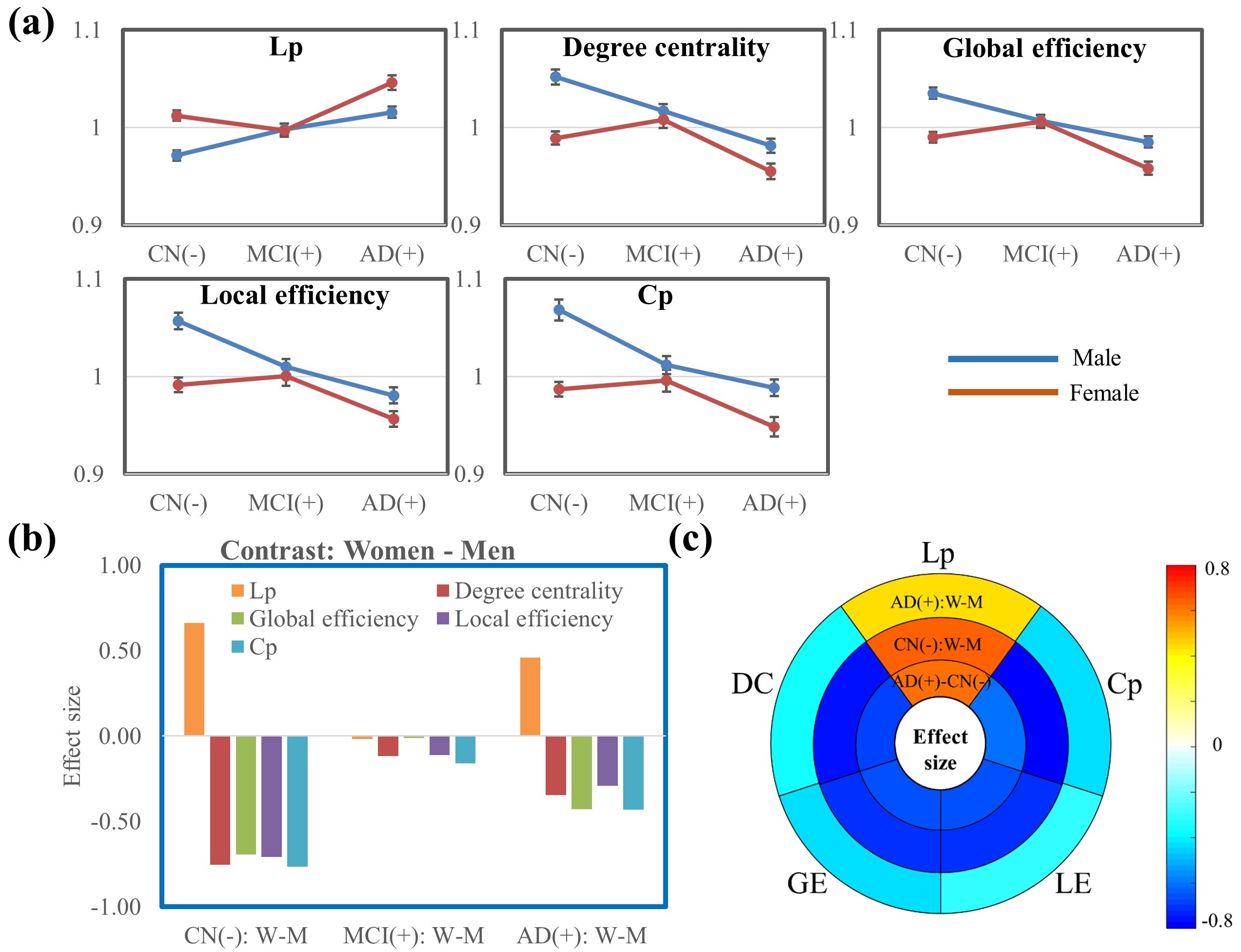
Figure 1. Graph
theory analysis at the global level. (a). Curves of the five global network
metrics from CN to MCI towards AD dementia. (b). Sex difference in CN, MCI, and
AD groups. (c). Effect size of the five global network metrics for the
contrasts AD dementia – CN (both women and men together), CN: Women – Men, and
AD dementia: Women – Men. The sex differences in both CN and AD dementia groups
are observed to be similar to the group differences between the AD dementia and
CN groups.
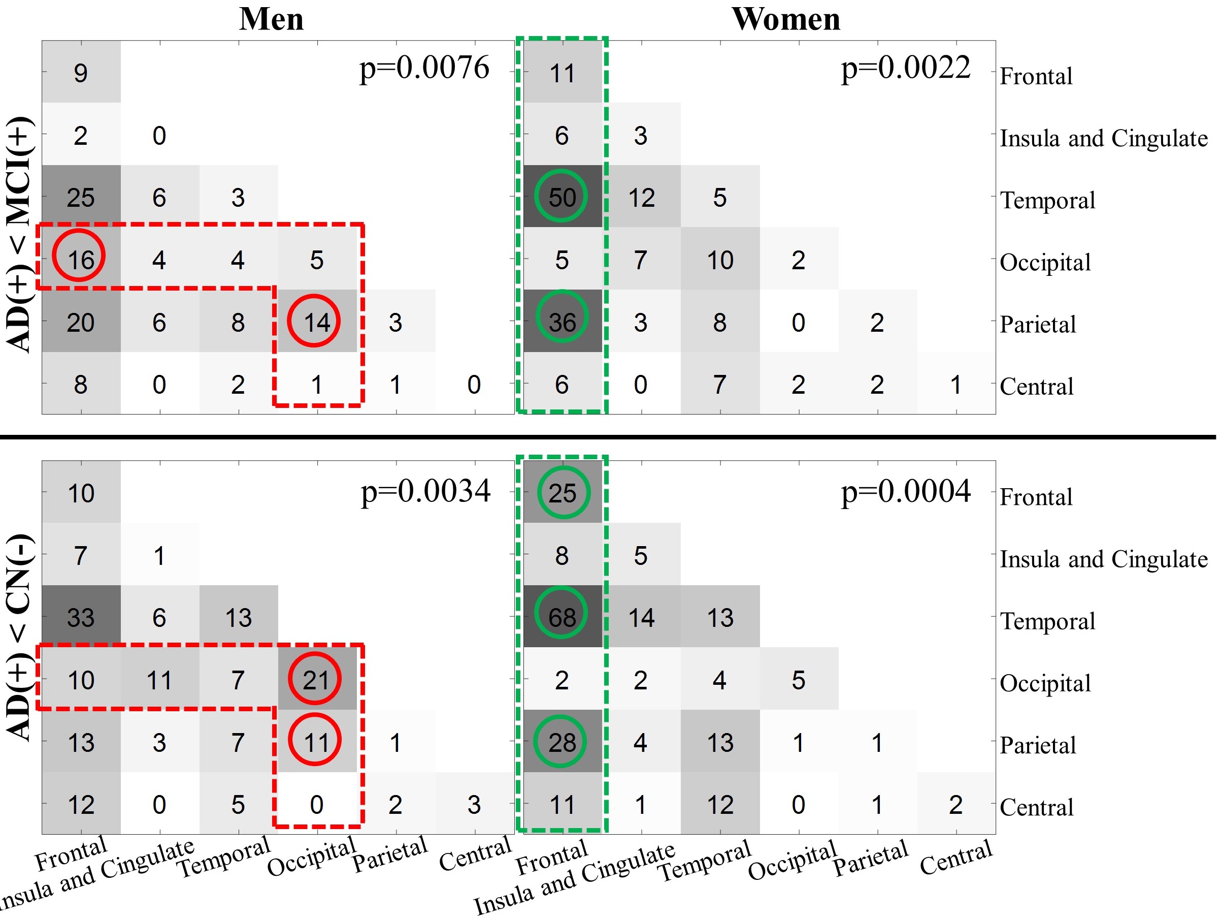
Figure
3. Summary of the edges in the significant clusters of contrast AD dementia
< MCI (top panel) and AD dementia < CN (bottom panel). Men and women
subjects are analyzed separately. The circles are used to mark the location
where women and men, for the same between-group contrast, have the difference
of the number of edges more than 10 (red: men>women; green: women>men).