huaguang yang1, zhi Wen1, Lanhua Hu1, Weiyin Vivian Liu2, Guoguang Fan3, and Yunfei Zha1
1Radiology, Renmin Hospital of Wuhan University, Wuhan, China, 2MR Research, GE Healthcare, Wuhan, China, 3The First Affiliated Hospital of China Medical University, Shenyang, China
1Radiology, Renmin Hospital of Wuhan University, Wuhan, China, 2MR Research, GE Healthcare, Wuhan, China, 3The First Affiliated Hospital of China Medical University, Shenyang, China
MSA patients with depression mainly showed the changes of central hub and functional connectivity mainly occurred in temporal lobe and subcortical thalamic nuclei of MSA patients with depression compared to those only with MSA
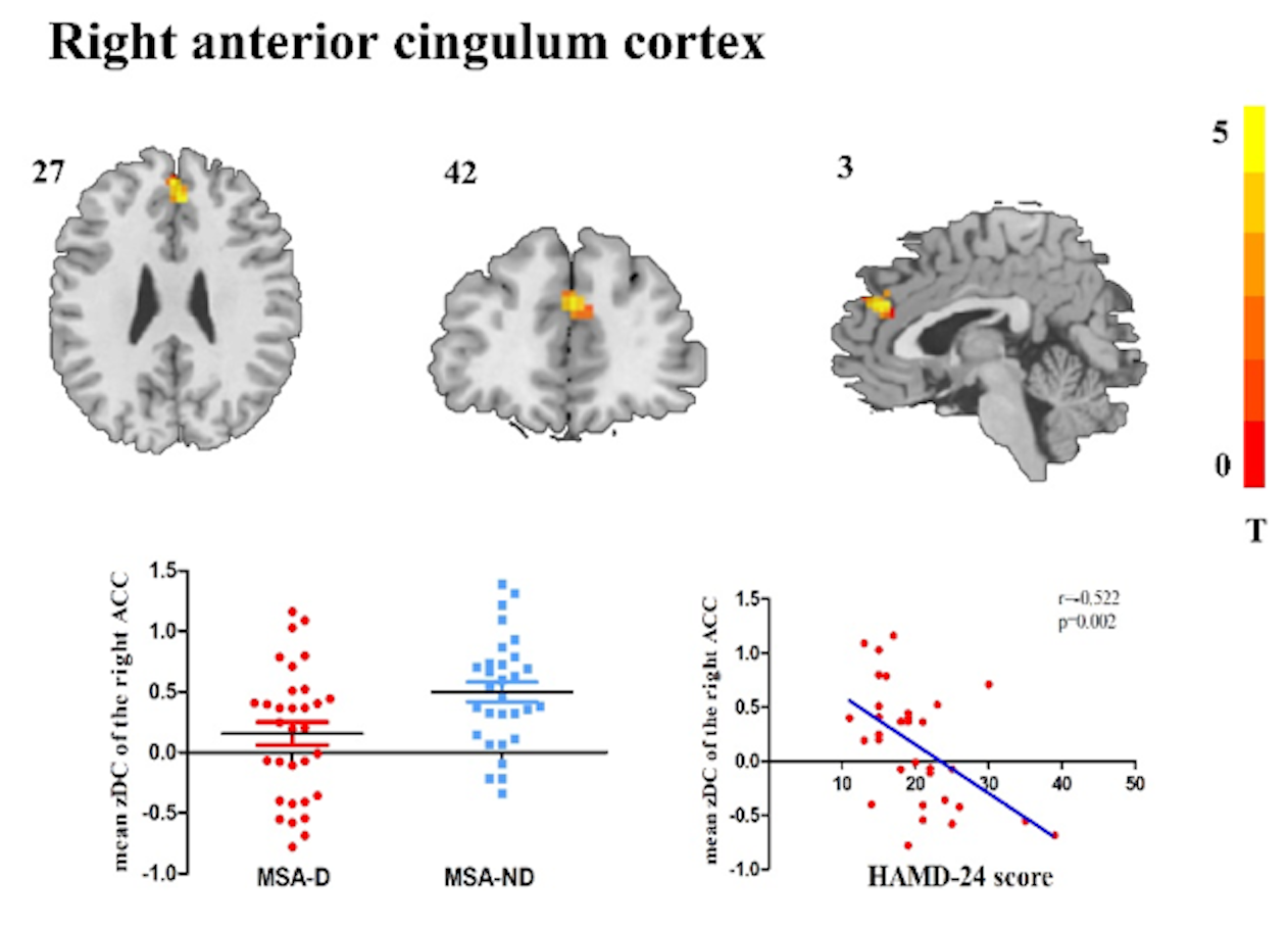
Figure 1. Compared with MSA-ND group, no difference of mean DC between groups was found. DC alteration was only found in the right ACC and correlated with clinical depression scores. Scatter plot showed a negative correlation between HAMD-24 scores and the right ACC zDC values in the MSA-D patients.
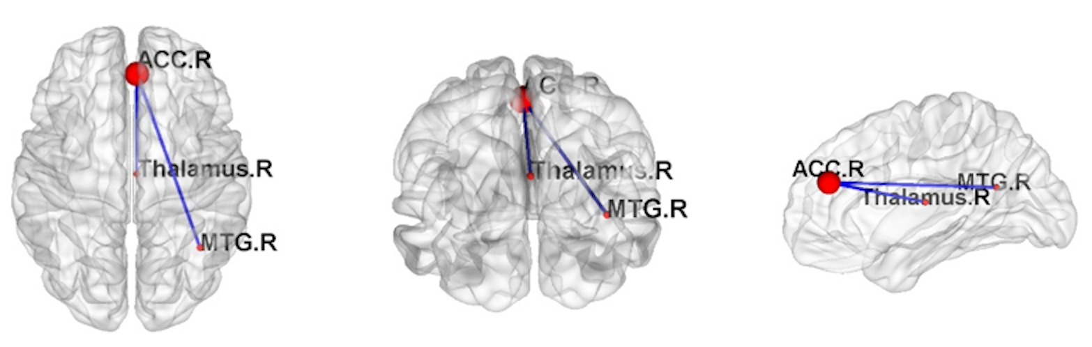
Figure 2. Post hoc two-sample t-test results of right ACC-seeded FC analyses between MSA-D and MSA-ND groups. The graph of the network is made by BrainNet (https://www.nitrc.org/projects/bnv/) software. Red node represents the node of brain area, and the blue edge represents the decreased functional connection. ACC.R, right anterior cingulate cortex; Thalamus.R, right thalamus; MTG.R,right middle temporal gyrus.