Elizabeth G. Keeling1,2, Maurizio Bergamino1, Lori Steffes1, Anna Burke3, and Ashley M. Stokes1
1Division of Neuroimaging Research, Barrow Neurological Institute, Phoenix, AZ, United States, 2School of Life Sciences, Arizona State University, Tempe, AZ, United States, 3Division of Neurology, Barrow Neurological Institute, Phoenix, AZ, United States
1Division of Neuroimaging Research, Barrow Neurological Institute, Phoenix, AZ, United States, 2School of Life Sciences, Arizona State University, Tempe, AZ, United States, 3Division of Neurology, Barrow Neurological Institute, Phoenix, AZ, United States
Compared to standard fMRI, SAGE-fMRI enables more robust distinction between healthy control and cognitively impaired groups through more sensitive assessment of AD-relevant brain regions via reduced susceptibility-induced signal dropout near air-tissue interfaces.
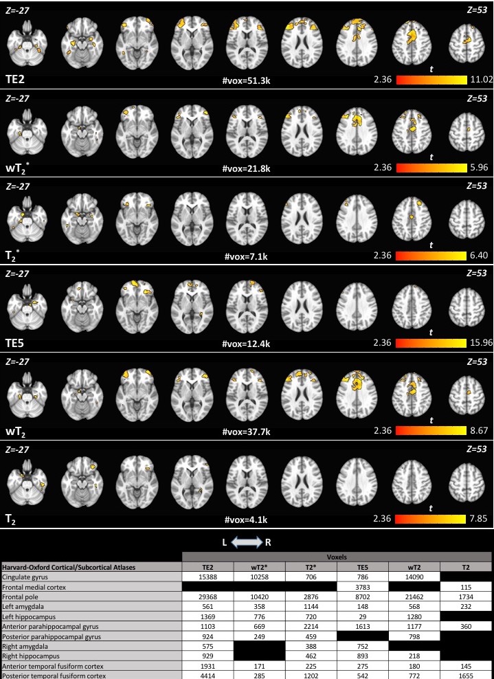
Significant differences in memory encoding task-related activation between HC and CI groups (HC>CI, p<0.05, cluster size>100) and voxel count in regions of interest for each SAGE-fMRI analysis method (GRE: TE2, T2*, wT2*; SE: TE5, T2, wT2).

Significant differences in memory recall task-related activation between HC and CI groups (HC>CI, p<0.05, cluster size>100) and voxel count in regions of interest for each SAGE-fMRI analysis method (GRE: TE2, T2*, wT2*; SE: TE5, T2, wT2).