Mei-Yu Yeh1,2, Ping Hou2, and Ho-Ling Liu 2
1Department of Biomedical Engineering and Environmental Sciences, National Tsing Hua University, Hsinchu, Taiwan, 2Department of Imaging Physics, The University of Texas MD Anderson Cancer Center, Houston, Houston, TX, United States
1Department of Biomedical Engineering and Environmental Sciences, National Tsing Hua University, Hsinchu, Taiwan, 2Department of Imaging Physics, The University of Texas MD Anderson Cancer Center, Houston, Houston, TX, United States
The proposed data-driven approach that does not require physiological monitoring during the BH MRI scan have a good agreement with the conventional method.
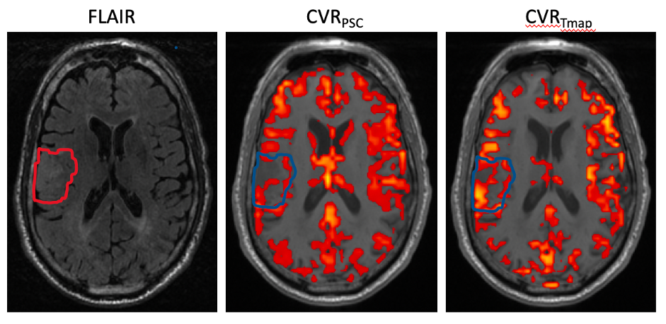
Fig.3 Image example from single patient. Left: FLAIR T2 image, middle: CVR maps from conventional BH, and the threshold was set to 0.35 for blood oxygen level dependent PSC. Right: CVR maps from the proposed method, and the threshold was set to t>3.45.
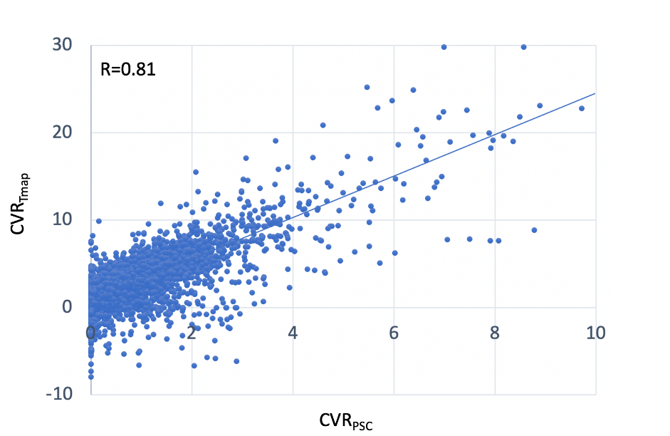
Fig.2 Voxel by voxel scatter plot between the two maps(CVRpsc and CVRTmap )