Charit Tippareddy1, Walter Zhao2, Andrew Sloan3,4, Jeffrey Sunshine5, Jill Barnholtz-Sloan6, Mark Griswold2,5, Dan Ma2,5, and Chaitra Badve5
1Case Western Reserve University School of Medicine, Cleveland, OH, United States, 2Department of Biomedical Engineering, Case Western Reserve University, Cleveland, OH, United States, 3Departments of Neurosurgery and Pathology, University Hospitals Cleveland Medical Center, Cleveland, OH, United States, 4Seidman Cancer Center and Case Comprehensive Cancer Center, Cleveland, OH, United States, 5Department of Radiology, University Hospitals Cleveland Medical Center, Cleveland, OH, United States, 6Department of Population and Quantitative Health Sciences, University Hospitals Cleveland Medical Center, Cleveland, OH, United States
1Case Western Reserve University School of Medicine, Cleveland, OH, United States, 2Department of Biomedical Engineering, Case Western Reserve University, Cleveland, OH, United States, 3Departments of Neurosurgery and Pathology, University Hospitals Cleveland Medical Center, Cleveland, OH, United States, 4Seidman Cancer Center and Case Comprehensive Cancer Center, Cleveland, OH, United States, 5Department of Radiology, University Hospitals Cleveland Medical Center, Cleveland, OH, United States, 6Department of Population and Quantitative Health Sciences, University Hospitals Cleveland Medical Center, Cleveland, OH, United States
Post contrast MRF T1, T2 maps allow quantitative
characterization of NET region in GBMs and METs. MRF-radiomics signatures can
differentiate NET regions of GBMs from METS and demonstrate unique differences
between near (within 1 cm of enhancing tumor) and far (beyond 1 cm) NET regions.
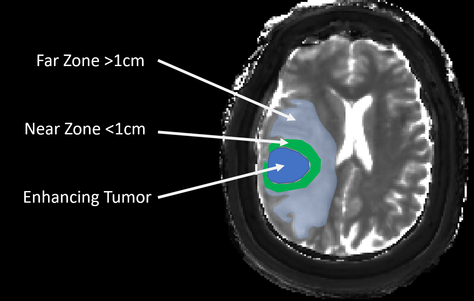
Zone analysis demonstrated on MRF T1 map in a right temporal GBM
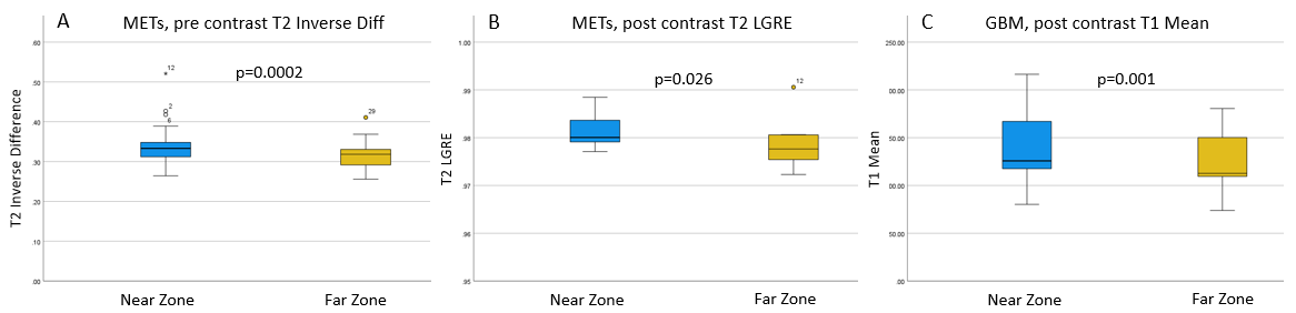
Boxplots of significant pre and post contrast
texture
features from NET
regions showing
near vs far zones in METs and GBMs