Simran Kukran1,2, Joely Smith3,4, Luke Dixon1,3, Ben Statton5, Sarah Cardona3, Lillie Pakzad-Shahabi6,7, Matthew Williams6,8, Dow-Mu Koh2,9, Rebecca Quest3,4, Matthew Orton2, and Matthew Grech-Sollars1,3
1Department of Surgery and Cancer, Imperial College London, London, United Kingdom, 2Department of Radiotherapy and Imaging, Institute of Cancer Research, London, United Kingdom, 3Department of Imaging, Imperial College Healthcare NHS Trust, London, United Kingdom, 4Department of Bioengineering, Imperial College London, London, United Kingdom, 5Medical Research Council, London Institute of Medical Sciences, Imperial College London, London, United Kingdom, 6Computational Oncology group, Institute for Global Health Innovation, Imperial College London, London, United Kingdom, 7John Fulcher Neuro-oncology Laboratory, Department of Brain Sciences, Imperial College London, London, United Kingdom, 8Radiotherapy Department, Charing Cross Hospital, London, United Kingdom, 9Department of Radiology, Royal Marsden Hospital, London, United Kingdom
1Department of Surgery and Cancer, Imperial College London, London, United Kingdom, 2Department of Radiotherapy and Imaging, Institute of Cancer Research, London, United Kingdom, 3Department of Imaging, Imperial College Healthcare NHS Trust, London, United Kingdom, 4Department of Bioengineering, Imperial College London, London, United Kingdom, 5Medical Research Council, London Institute of Medical Sciences, Imperial College London, London, United Kingdom, 6Computational Oncology group, Institute for Global Health Innovation, Imperial College London, London, United Kingdom, 7John Fulcher Neuro-oncology Laboratory, Department of Brain Sciences, Imperial College London, London, United Kingdom, 8Radiotherapy Department, Charing Cross Hospital, London, United Kingdom, 9Department of Radiology, Royal Marsden Hospital, London, United Kingdom
MRF was found to give highly repeatable T1 and T2 measurements that were strongly and significantly correlated to measurements from established mapping techniques in both tumour and normal appearing contralateral brain tissue of glioma patients.
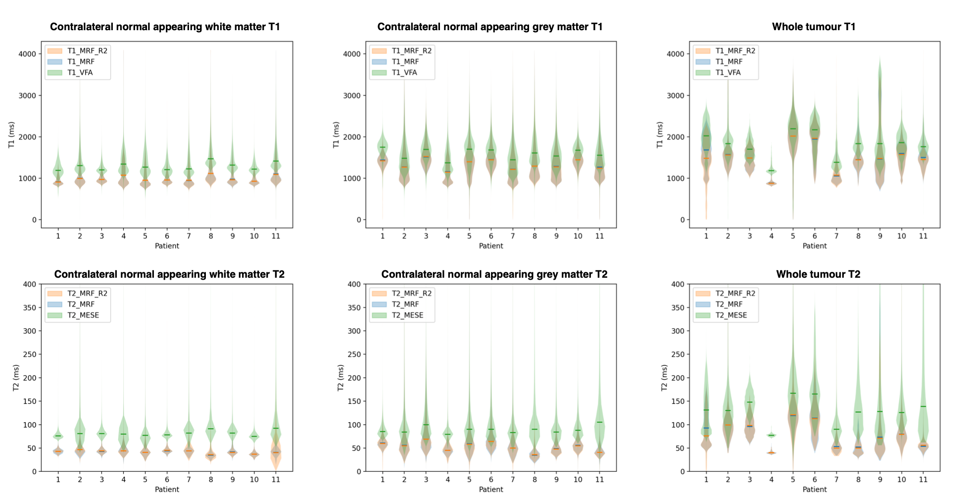
Violin plots with medians for T1 and T2 values of NAC GM, NAC WM, and tumour regions from repeated MRF and established T1 VFA and T2 MESE relaxometry. MRF repeat violins (red/blue) are almost indistinguishable, indicating MRF measurements are highly repeatable. MRF values are consistently lower than established techniques (green).
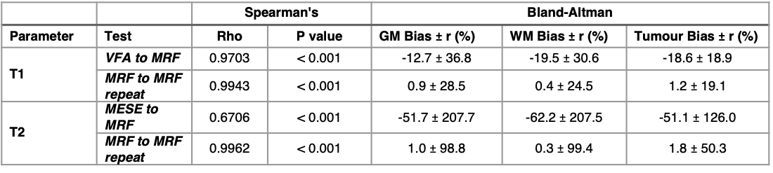
Table 2: Summary of Spearman’s Rho correlation testing and Bland-Altman statistics for: VFA vs. MRF T1, MESE vs. MRF T2 and repeated MRF measurements of both T1 and T2.