Andrew Mao1,2,3, Sebastian Flassbeck1,2, Cem Gultekin4, Xiaoxia Zhang1,2, and Jakob Asslaender1,2
1Center for Biomedical Imaging, Department of Radiology, New York University School of Medicine, New York, NY, United States, 2Center for Advanced Imaging Innovation and Research, New York University School of Medicine, New York, NY, United States, 3Vilcek Institute of Graduate Biomedical Sciences, New York University School of Medicine, New York, NY, United States, 4Courant Institute of Mathematical Sciences, New York University, New York, NY, United States
1Center for Biomedical Imaging, Department of Radiology, New York University School of Medicine, New York, NY, United States, 2Center for Advanced Imaging Innovation and Research, New York University School of Medicine, New York, NY, United States, 3Vilcek Institute of Graduate Biomedical Sciences, New York University School of Medicine, New York, NY, United States, 4Courant Institute of Mathematical Sciences, New York University, New York, NY, United States
We optimize an MR fingerprinting sequence for SNR efficiency using hybrid-state theory, and apply it in vivo demonstrating simultaneous quantification of myelin water fraction, T1 and T2 of axonal/extra-axonal water in the full brain with high spatial resolution with a 14 minute scan.
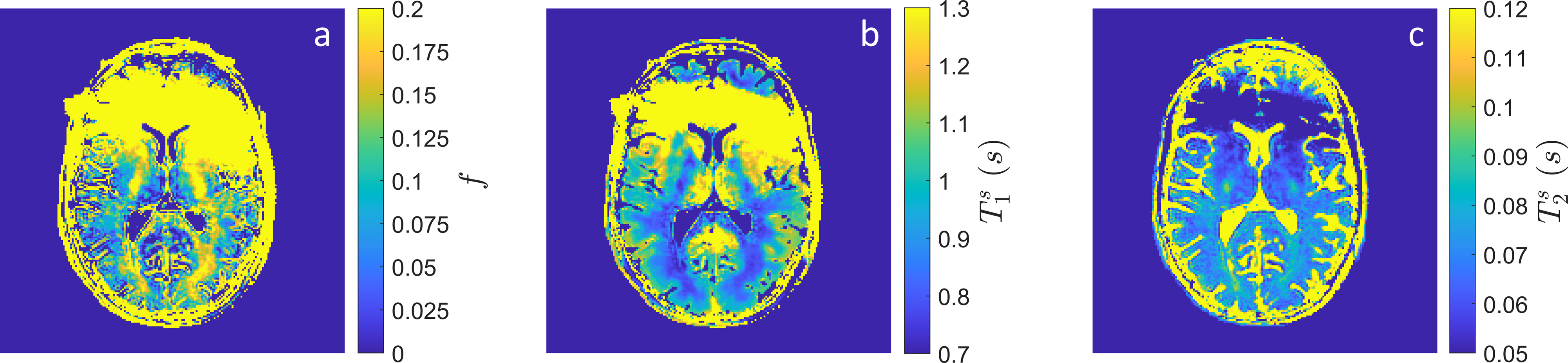
Figure 4: In vivo quantitative maps of (a) $$$f$$$, (b) $$$T_1^s$$$ and (c) $$$T_2^s$$$ from a single axial slice through the brain at the level of the lateral ventricles. The fast compartment ($$$T_1^f$$$ and $$$T_2^f$$$) was fixed to literature values for the fitting process. Note that the anterior brain is corrupted by $$$B_0$$$ artifact arising from metal in the volunteer's face mask.
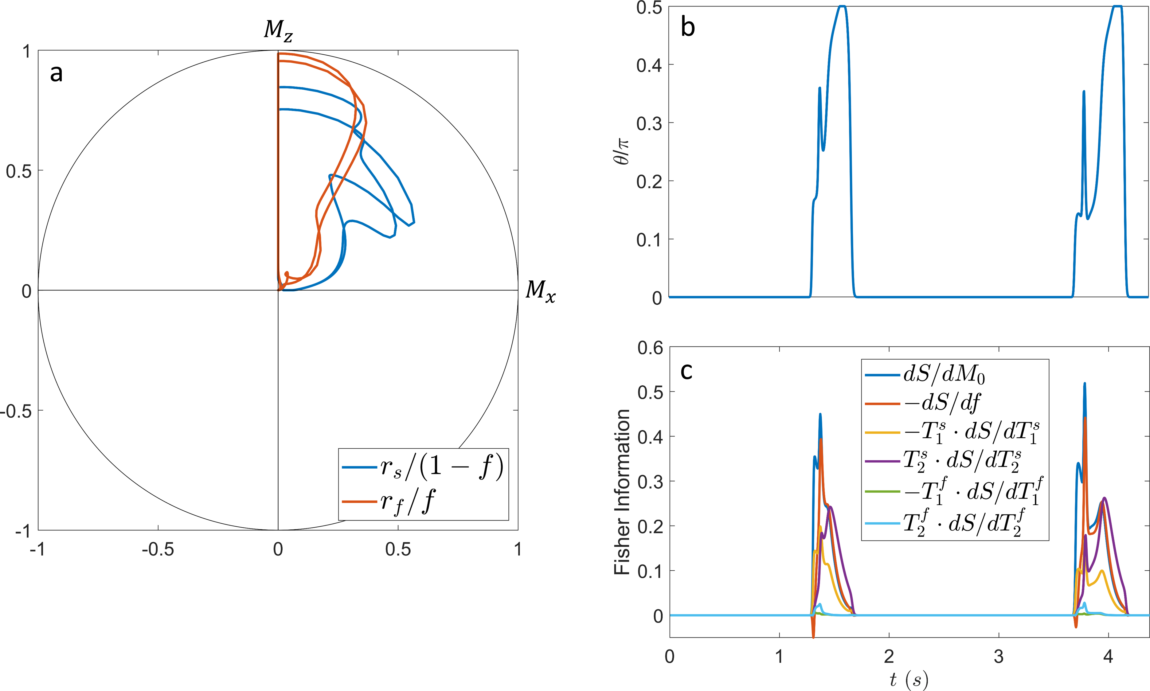
Figure 2: (a) Spin dynamics of the slow (blue) and fast (red) compartment on the Bloch sphere. Note that the magnetization of each compartment is normalized by their respective fractions for visual clarity. (b) Numerically optimized $$$\vartheta$$$ pattern over time. (c) Fisher information over time. The optimized sequence prefers two distinct information dense periods per $$$T_{cyc}$$$.