Pavan Poojar1,2, Enlin Qian1, and Sairam Geethanath 1,2
1Columbia Magnetic Resonance Research Center, Columbia University, New York, NY, United States, 2Dayananda Sagar College of Engineering, Bangalore, India
1Columbia Magnetic Resonance Research Center, Columbia University, New York, NY, United States, 2Dayananda Sagar College of Engineering, Bangalore, India
We improved our tailored MRF implementation by adding two contrasts (water and fat), reducing scan time by 25% (from 5:27(min:sec) to 4:07(min:sec)) and reducing reconstruction time (from ~40mins to ~3mins). TMRF SNR and contrast was better than MRF.
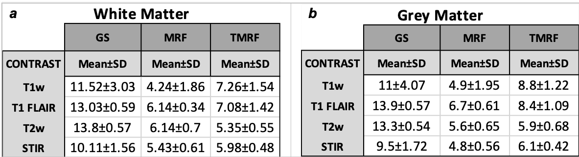
Table
2: Mean
and standard deviation (SD) of signal to noise ratio (SNR) for (a)
white matter
(WM)
and (b) grey matter (GM). The SNR was calculated for all the three methods –
GS, MRF and TMRF (columns) and for all the four contrasts (rows). Initially, WM
and GM were segmented and then SNR was calculated using 3D slicer software. SNR
was measured using “difference image” method, where the same brain images were
acquired twice with identical conditions. The SNR for GS>TMRF>MRF for WM
and GM for all the contrasts.
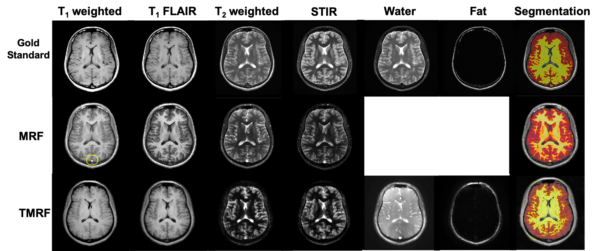
Figure 1: Qualitative
healthy brain images obtained using gold standard method (first row), magnetic
resonance fingerprinting (MRF) (second
row) and tailored
MRF
(TMRF) (third
row).
The color
axis bar for each image is different. Each
column
represents different contrasts along
with the representative grey matter and white matter segmentation (last column)
using 3D slicer. Images obtained using MRF method were synthetically generated
and have flow artifacts as shown in the yellow circle.