Gastao Cruz1, Carlos Velasco1, Olivier Jaubert1, Haikun Qi1, René M. Botnar1, and Claudia Prieto1
1School of Biomedical Engineering and Imaging Sciences, King's College London, London, United Kingdom
1School of Biomedical Engineering and Imaging Sciences, King's College London, London, United Kingdom
ECG-triggered, multi-echo gradient-echo cardiac MRF using large
acquisition window is proposed to simultaneously map T1, T2, T2* and fat
fraction. A low-rank non-rigid motion correction reconstruction corrects cardiac
motion, yielding 4 co-registered parameter maps from a single scan.
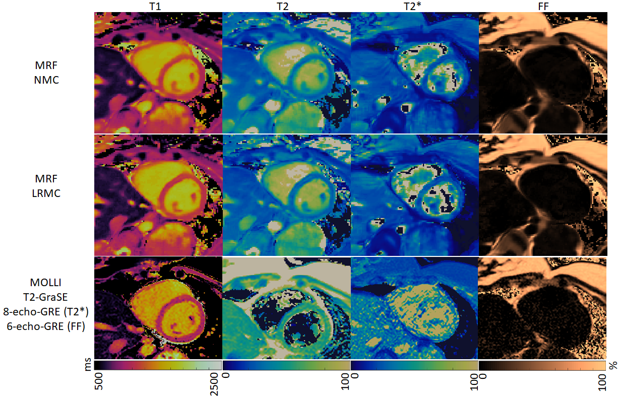
Fig.3
T1, T2, T2* and FF maps for one representative subject obtained with (long
cardiac acquisition window) MRF with no motion correction (NMC), with motion correction
(proposed LRMC) and the corresponding conventional methods (MOLLI, T2-GraSE,
8-echo GRE for T2* and 6-echo GRE for FF). Residual artefacts (predominantly
blurring) are present with NMC and are reduced with LRMC, achieving comparable
quality to conventional single-parameter mapping methods.
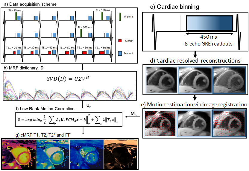
Fig.1
Diagram for the proposed T1, T2, T2* and FF cardiac MRF. a) ECG-triggered
data is acquired with varying preparation pulses; b) low rank subspace
is estimated from the MRF dictionary; c) long acquisition window data is
binned into multiple cardiac phases; d) cardiac resolved images are
reconstructed with LRI-HDPROST; e) motion is estimated with free-form
deformations; f) LRMC is performed producing a set of motion corrected
singular values for all echoes; g) T1 and T2 is estimated via MRF
dictionary matching, T2* and FF are estimated via a water/fat separation algorithm.