Telly Ploem1, Jaap Boon1, Ingo Hermann1,2, Cole S. Simpson3, Joe F Juffermans4, Tom M. Piscaer5, Hildo J Lamb4, Nazli Tümer6, Joao Tourais1, and Sebastian Weingärtner1
1Magnetic Resonance Systems Lab, Department of Imaging Physics, Delft University of Technology, Delft, Netherlands, 2Computer Assisted Clinical Medicine, Medical Faculty Mannheim, Heidelberg University, Mannheim, Germany, 3Department of Mechanical Engineering, Stanford University, Stanford, CA, United States, 4Department of Radiology, Leiden University Medical Center, Leiden, Netherlands, 5Orthopaedic Surgery, Erasmus University Medical Centre, Rotterdam, Netherlands, 6Department of Biomechanical Engineering, Delft University of Technology, Delft, Netherlands
1Magnetic Resonance Systems Lab, Department of Imaging Physics, Delft University of Technology, Delft, Netherlands, 2Computer Assisted Clinical Medicine, Medical Faculty Mannheim, Heidelberg University, Mannheim, Germany, 3Department of Mechanical Engineering, Stanford University, Stanford, CA, United States, 4Department of Radiology, Leiden University Medical Center, Leiden, Netherlands, 5Orthopaedic Surgery, Erasmus University Medical Centre, Rotterdam, Netherlands, 6Department of Biomechanical Engineering, Delft University of Technology, Delft, Netherlands
MRF-EPI enables whole knee quantification of T1 and T2* across 16 slices in 3
minutes. Phantom scans demonstrate good agreement with reference methods and visually high map quality is achieved in vivo.
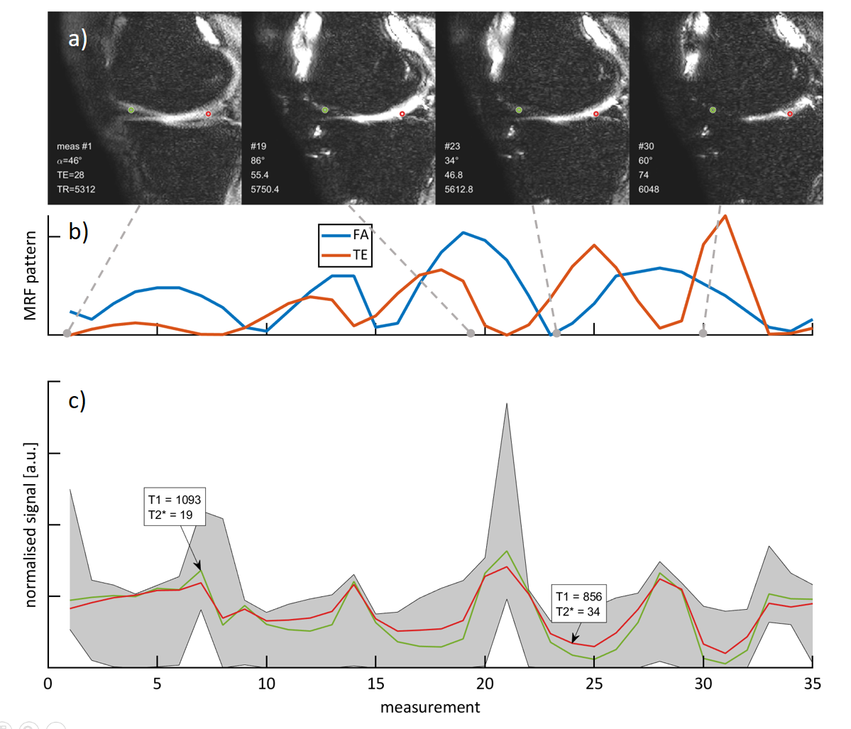
Figure 1: Schematic overview of the MRF-EPI sequence. a) Baseline images for
four different measurements showing contrast variations throughout the MRF
acquisition. Gray dashed lines point to the parameters used for acquiring the
image. The red and green circles indicate the locations of the fingerprints shown
in c). b) Flip angle (α) (34 to 84 degree) and echo time (TE) (28 to 89 ms) pattern of the MRF-EPI sequence. c) Signal evolution for 2 different fingerprints in
the cartilage of a healthy subject, with the corresponding range of the complete
dictionary (gray shading).
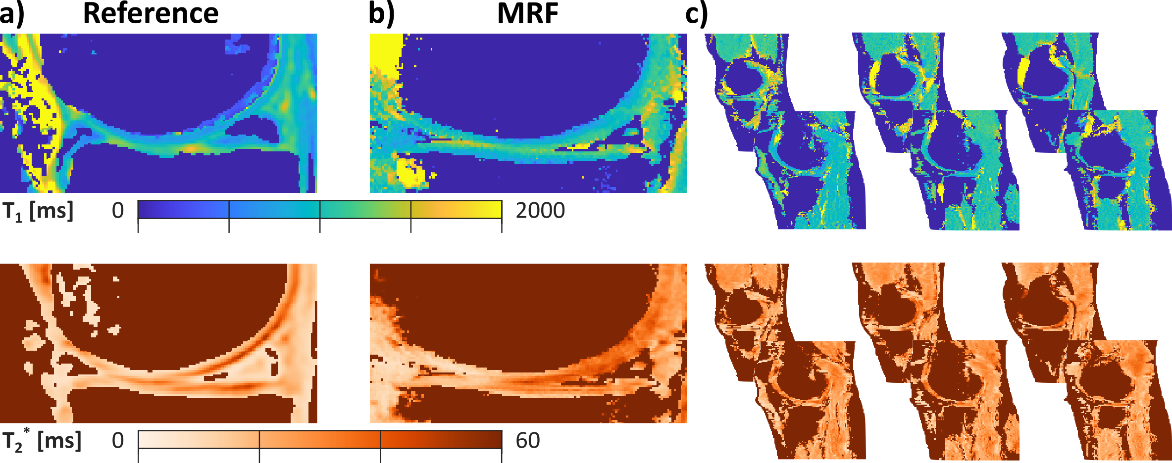
Figure 5: In vivo T1 and T2* maps for one slice of MRF (a) and reference scans
(MOLLI and multi GRE) (b). c) multiple MRF maps illustrating full medial
and lateral coverage with the proposed whole-knee technique.