Christian Guenthner1, Peter Koken2, Peter Boernert2,3, and Sebastian Kozerke1
1University and ETH Zurich, Zurich, Switzerland, 2Philips Research, Hamburg, Germany, 3Leiden University Medical Center, Leiden, Netherlands
1University and ETH Zurich, Zurich, Switzerland, 2Philips Research, Hamburg, Germany, 3Leiden University Medical Center, Leiden, Netherlands
The feasibility of concurrent water/fat separation
and T1/T2 mapping using FISP-MR Fingerprinting was assessed on a lower-field
0.75T MRI.
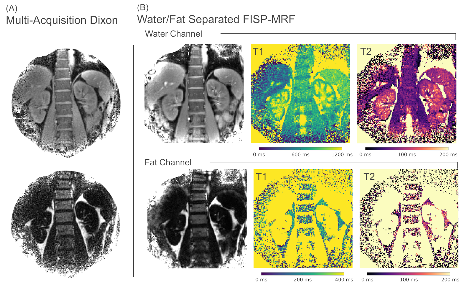
Figure
4: (A) Reference cartesian Dixon scan and (B) water/fat
resolved MRF. Abdominal water/fat separated FISP-MRF results show proton
density and T1/T2 parameter maps for both fat and water separately. A mask was
applied to the parameter maps to only show regions of sufficient proton signal.
The proton density images compare well between MRF and the classical Dixon
sequence, with inflow effects being present in the MRF scan leading to bright
vessels.
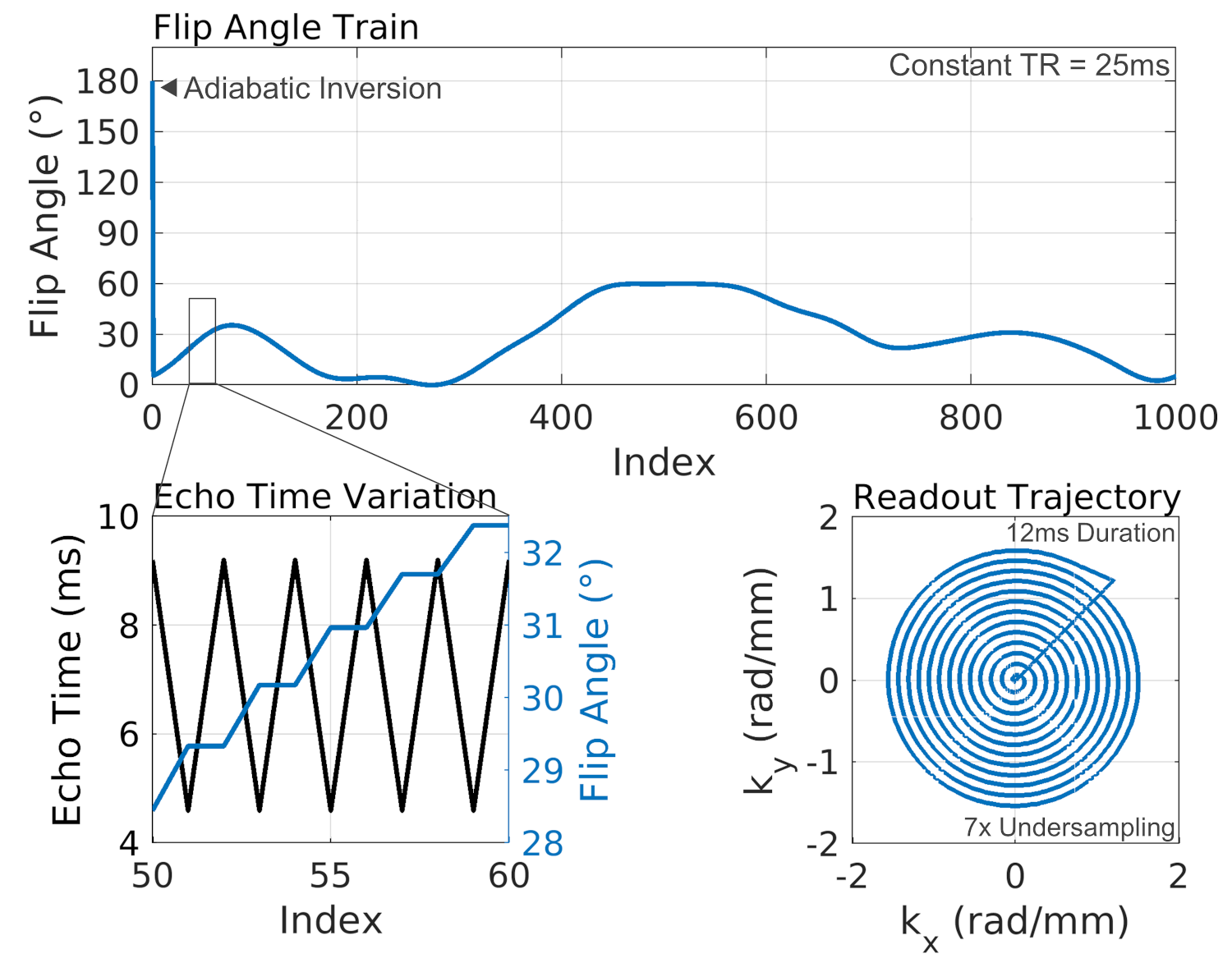
Figure
1: FISP-MRF sequence with interleaved in- and
out-of-phase echo times for water/fat separation at 0.75T. For each flip angle,
an out-of-phase image is acquired first with an echo time of 4.6 ms
followed by an in-phase image with 9.21 ms echo time (N.B.: the water/fat
shift at 0.75T is approximately 108 Hz). For acquisition, a 12 ms Archimedean
spiral readout with 7 interleaves and 2mm nominal resolution was used. For each
time point only one spiral interleave was acquired, leading to a 7-fold
undersampled acquisition