Ye Li1,2, Zidong Wei1,2,3, Shihong Han3, Shuheng Zhang3, Qiang He3, Xiaoliang Zhang4, Xin Liu1,2, and Hairong Zheng1,2
1Lauterbur Imaging Research Center, Shenzhen Institutes of Advanced Technology, Chinese Academy of Sciences, Shenzhen, China, 2Key Laboratory for Magnetic Resonance and Multimodality Imaging of Guangdong Province, Shenzhen, China, 3United Imaging Healthcare, Shanghai, China, 4Department of Biomedical Engineering, State University of New York at Buffalo, NY, NY, United States
1Lauterbur Imaging Research Center, Shenzhen Institutes of Advanced Technology, Chinese Academy of Sciences, Shenzhen, China, 2Key Laboratory for Magnetic Resonance and Multimodality Imaging of Guangdong Province, Shenzhen, China, 3United Imaging Healthcare, Shanghai, China, 4Department of Biomedical Engineering, State University of New York at Buffalo, NY, NY, United States
The image quality at 5 T
was largely improved comparing with 3 T, which indicated 5 T scanner’s
potential in clinical and brain science applications.
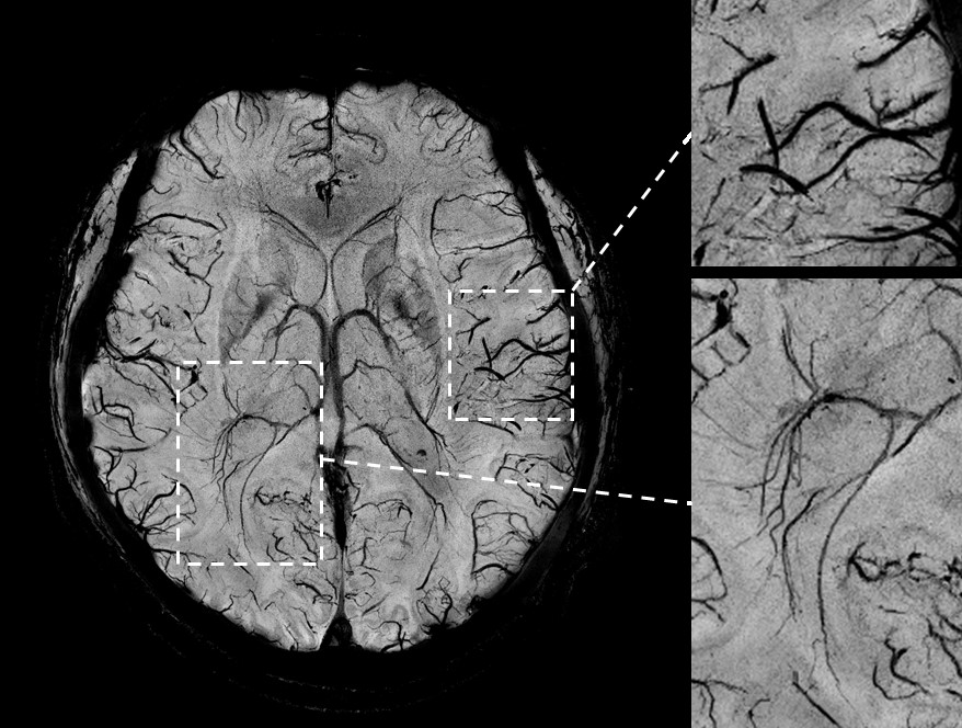
Figure 3: Susceptibility weighted image
(left) and zoomed in images (right) with 0.35*0.35*1.5 mm3 at 5T.
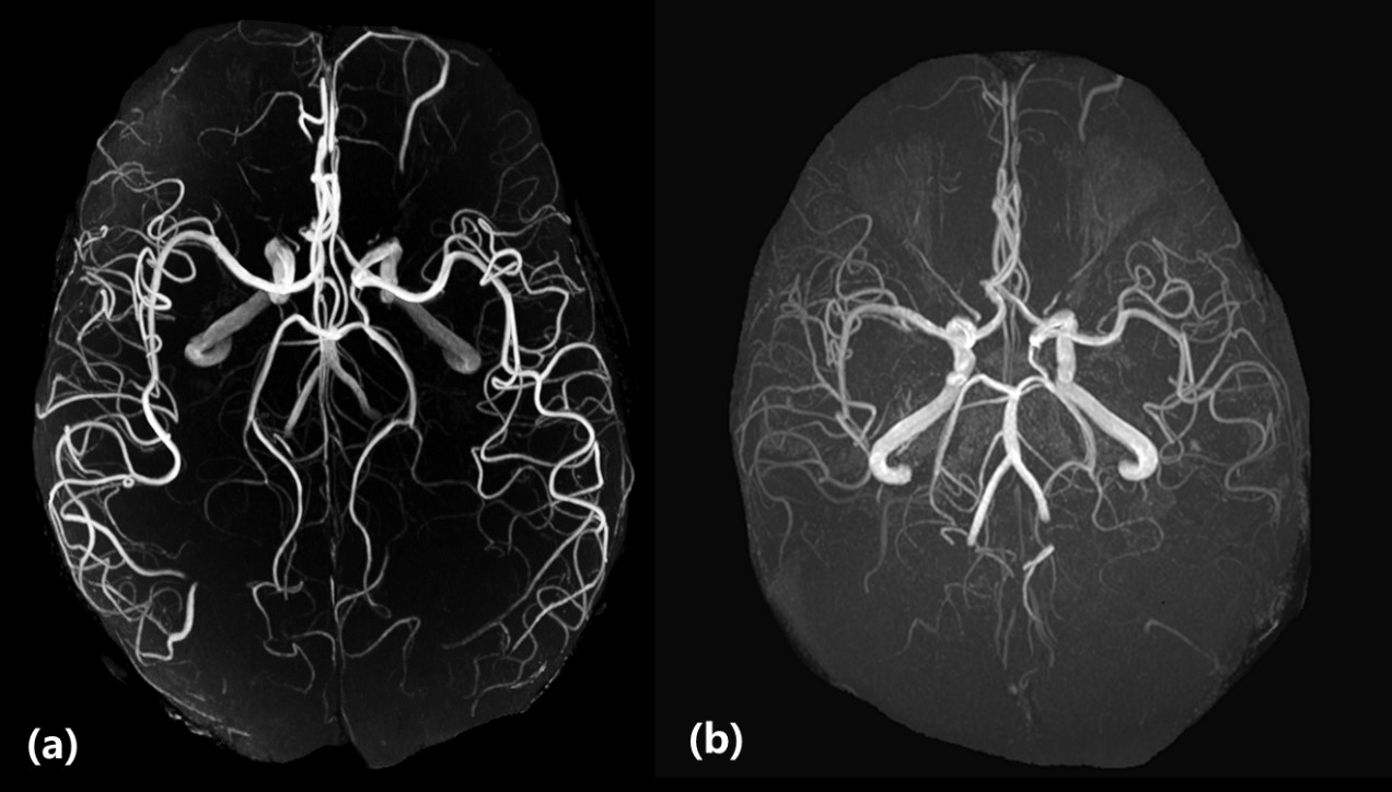
Figure
2: Magnetic resonance angiography images acquired by TOF sequence with 0.6*0.6*0.6
mm3 resolution. (a) and (b) were acquired at 5T and 3T respectively
using the same coils as in Figure 1.