Ibrahim A. Elabyad1, Maxim Terekhov1, Michael Hock1, David Lohr1, and Laura M. Schreiber1
1Chair of Molecular and Cellular Imaging, Comprehensive Heart Failure Center (CHFC), University Hospital Wuerzburg, Wuerzburg, Germany
1Chair of Molecular and Cellular Imaging, Comprehensive Heart Failure Center (CHFC), University Hospital Wuerzburg, Wuerzburg, Germany
The 16-element antisymmetric dipole array fits with the same dimensions as
an 8-element straight dipole array. Anatomical T1-weighted images of the pig heart were acquired on a fresh cadaver (15min postmortem) at
high spatial-resolution (0.5×0.5×4mm3).
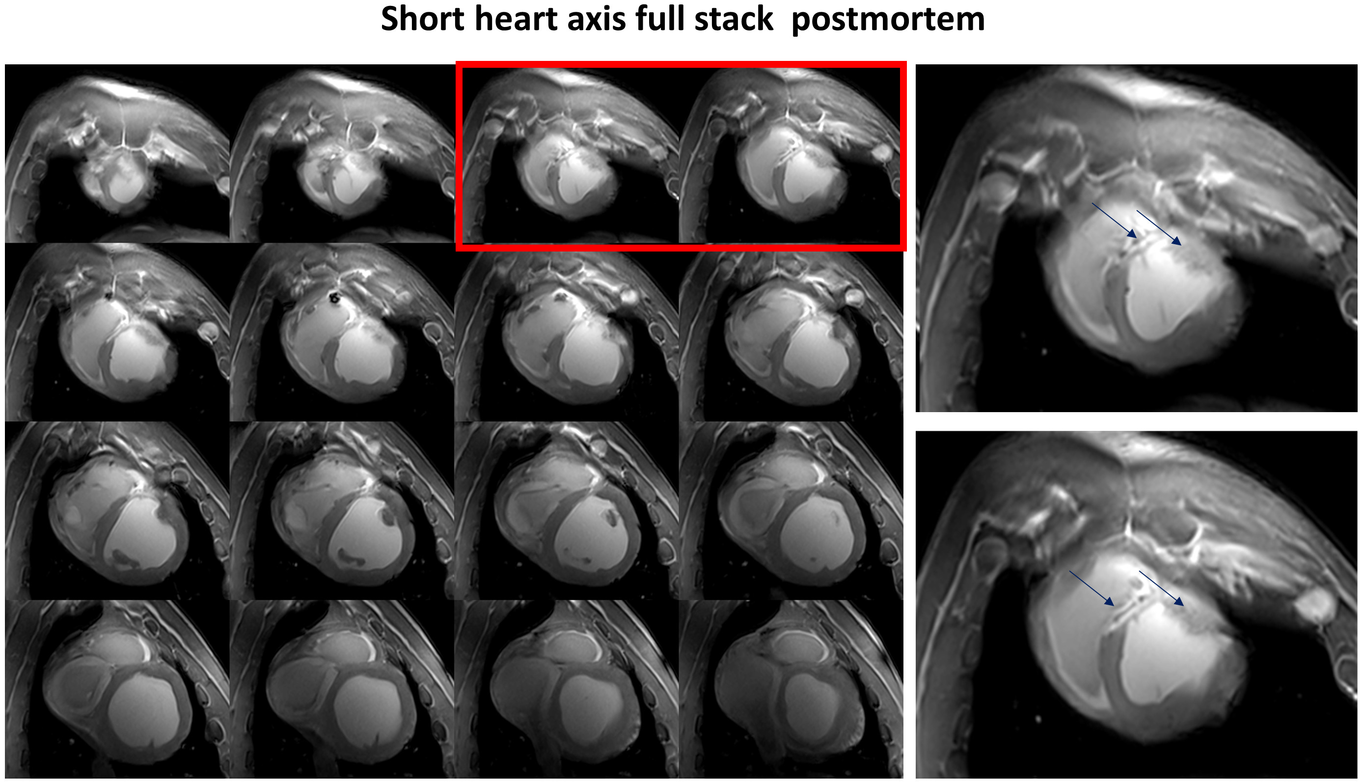
Figure 5. Slices of the short-axis
view stack of the pig heart acquired post-mortem. The right panels show
increased image slices marked red on the left panel. The LGE in the
post-infarction scar is observed in the septal and anterior wall (arrows).
Sufficient $$${B_1^+}$$$ field penetration and homogeneity without destructive interferences are
achieved in the whole heart in both anterior-posterior and basal-apical
directions.
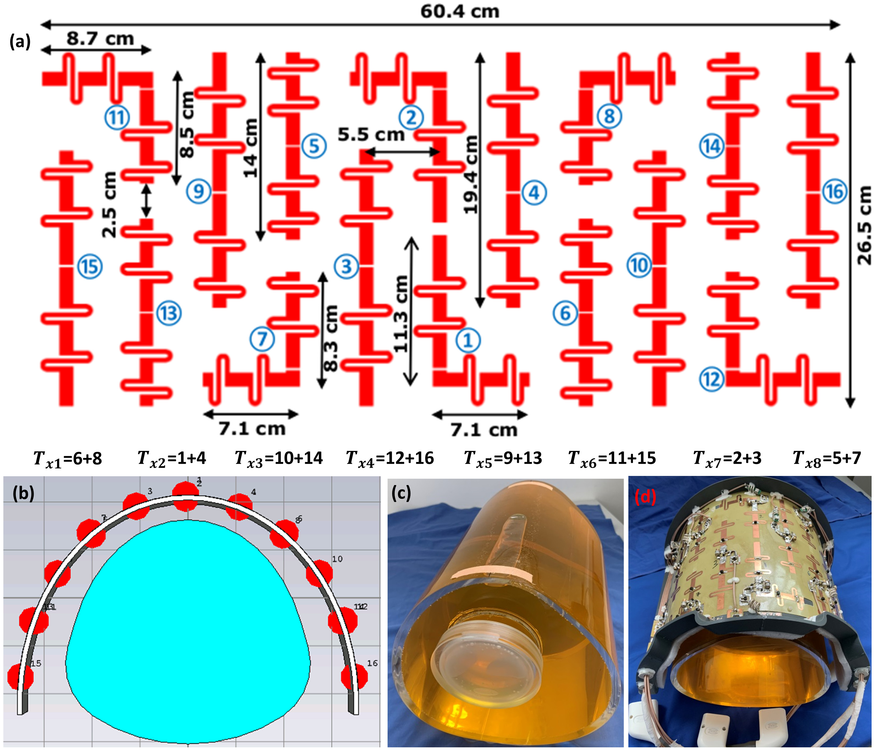
Figure 2. (a) Schematic
of the mono-surface antisymmetric 16-element dipole
antenna array with element dimensions,
paring channels, and channel numbers with every two neighboring dipoles to
form 8Tx-channels for the pTx system (8Tx/16Rx). (b)
RF simulation model as simulated in CST-MWS of the new dipole antenna array
loaded with a dedicated pig thorax phantom. (c)-(d)
Prototypes of the dedicated pig thorax phantom and the antisymmetric dipole antenna array.