Maxim Terekhov1, David Lohr1, Theresa Reiter2, Ibrahim A. Elabyad1, Michael Hock1, and Laura M. Schreiber1
1Chair of Molecular and Cellular Imaging, University Hospital Würzburg, Comprehensive Heart Failure Center, Wuerzburg, Germany, 2Department of Internal Medicine I, Cardiology, University Hospital Würzburg, Wuerzburg, Germany
1Chair of Molecular and Cellular Imaging, University Hospital Würzburg, Comprehensive Heart Failure Center, Wuerzburg, Germany, 2Department of Internal Medicine I, Cardiology, University Hospital Würzburg, Wuerzburg, Germany
We present the initial experience of using a commercial version of the 8Tx/16Rx thorax array operating in the pTX-Compatibility Mode of the scanner for cardiac MRI at 7T.
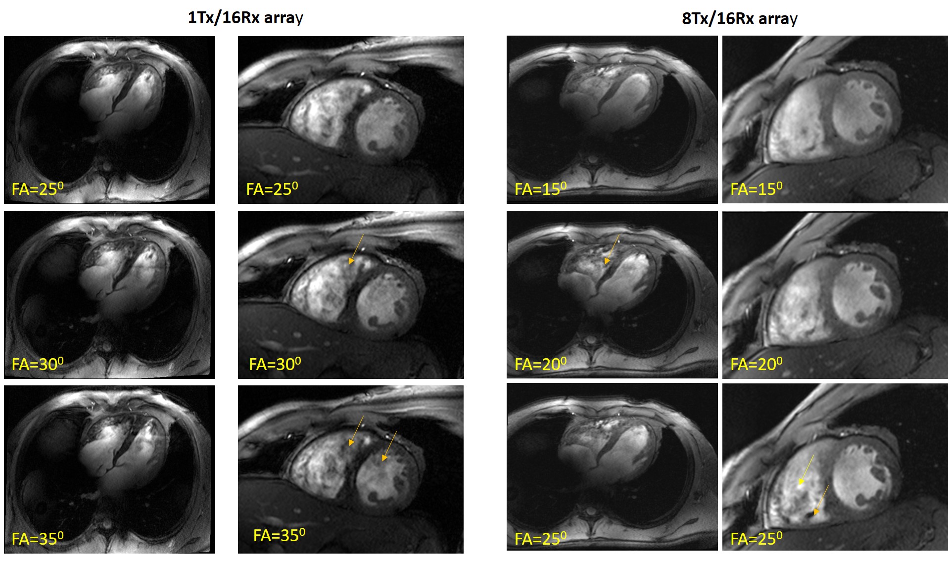
Figure 2 Long
and short-axis view of the heart acquired at different flip-angles with both
arrays. A pronounced appearance of blood-flow induced artifacts is observed
with the new array, probably due to lower B1-gradients in
the anterior-posterior direction.
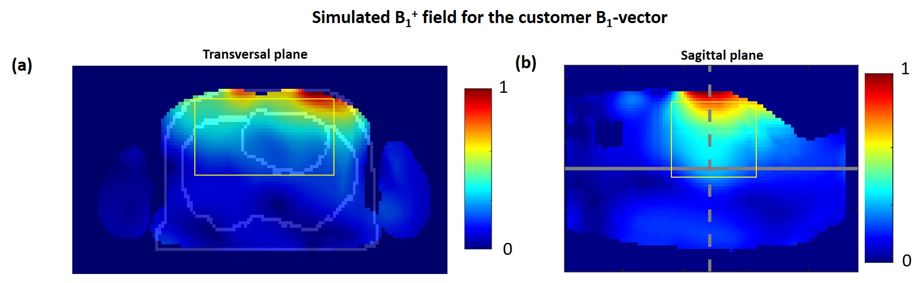
Figure 5 Result from optimized B1-vectors calculations. Panels (a) and (b) show the
B1-field on the "Duke" model in the transversal and sagittal slices
respectively. A sufficient penetration depth with better coverage of the
posterior heart (lung and heart contours are shown) is predicted by these
simulations for future measurements.