Jana Vincent1,2,3, Clyve Konrad Follante1, Ersin Bayram4, Lloyd Estkowski5, Ty Cashen6, Mark Giancola1, Victor Taracila1, Yun-Jeong Stickle1, Lalit Rai1, Venkata Malasani1, Nicole Wake7,8, Vichiry Yan1, Robert Stormont9, Joseph Rispoli2,10, and Fraser Robb1
1GE Healthcare Coils, Aurora, OH, United States, 2Weldon School of Biomedical Engineering, Purdue University, West Lafayette, IN, United States, 3Basic Medical Sciences, Purdue University, West Lafayette, IN, United States, 4Global MR Applications & Workflow, GE Healthcare, Houston, TX, United States, 5Global MR Applications & Workflow, GE Healthcare, Waukesha, WI, United States, 6Global MR Applications and Workflow, GE Healthcare, Madison, WI, United States, 7Albert Einstein College of Medicine, Montefiore Medical Center, Bronx, NY, United States, 8Center for Advanced Imaging Innovation and Research, Department of Radiology, NYU School of Medicine, New York, NY, United States, 9GE Healthcare, Waukesha, WI, United States, 10School of Electrical & Computer Engineering, Purdue University, West Lafayette, IN, United States
1GE Healthcare Coils, Aurora, OH, United States, 2Weldon School of Biomedical Engineering, Purdue University, West Lafayette, IN, United States, 3Basic Medical Sciences, Purdue University, West Lafayette, IN, United States, 4Global MR Applications & Workflow, GE Healthcare, Houston, TX, United States, 5Global MR Applications & Workflow, GE Healthcare, Waukesha, WI, United States, 6Global MR Applications and Workflow, GE Healthcare, Madison, WI, United States, 7Albert Einstein College of Medicine, Montefiore Medical Center, Bronx, NY, United States, 8Center for Advanced Imaging Innovation and Research, Department of Radiology, NYU School of Medicine, New York, NY, United States, 9GE Healthcare, Waukesha, WI, United States, 10School of Electrical & Computer Engineering, Purdue University, West Lafayette, IN, United States
Here we present the first 60-channel, high resolution, lightweight, flexible, supine breast coil. The coil mitigates respiratory and motion artifacts through
high acceleration which is made possible with a high channel count and
commensurate SNR that, in turn, facilitates supine imaging.
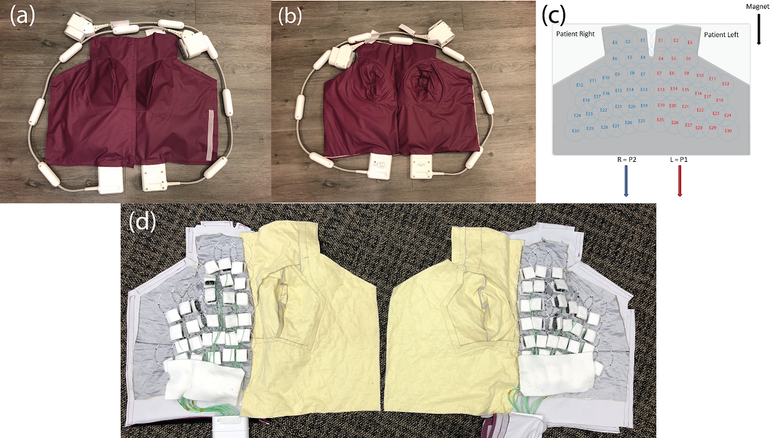
Figure 1. Complete breast/torso coil setup: (a) Front and (b) back mechanical assembly, (c-d) Coil assembly
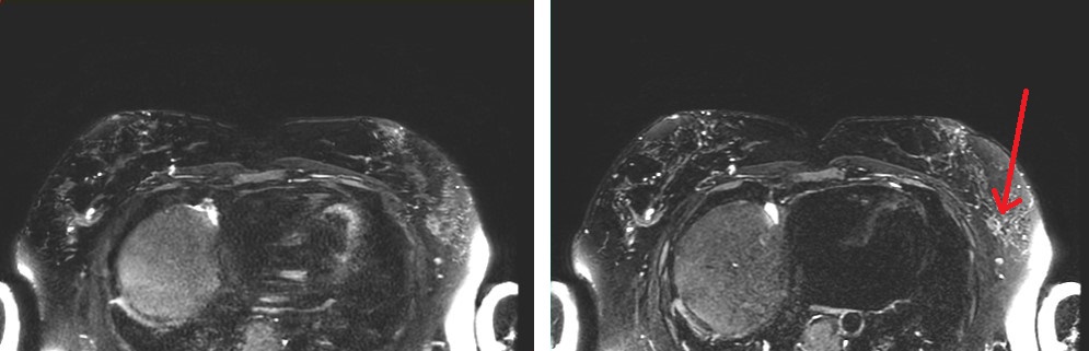
Figure 3. SSFSE,
Left: acceleration factor of 3, Right: acceleration factor of 4. Improved
sharpness is observed with higher acceleration due to reduced T2 blurring resulting
from a shortened echo train. Arrow indicates region of fibroglandular tissue.