Nikos Priovoulos1, Thomas Roos1, Ozlem Ipek2, Ettore Meliado3, Richard Nkrumah2, Dennis Klomp3, and Wietske van der Zwaag1
1Spinoza Center, Amsterdam, Netherlands, 2King’s College London, London, United Kingdom, 3University Medical Center Utrecht, Utrecht, Netherlands
1Spinoza Center, Amsterdam, Netherlands, 2King’s College London, London, United Kingdom, 3University Medical Center Utrecht, Utrecht, Netherlands
The combination of 32Rx dense surface receives with a dedicated 3Tx transmit results in increased B1+, SNR and BOLD sensitivity in the human cerebellum at 7Tesla.
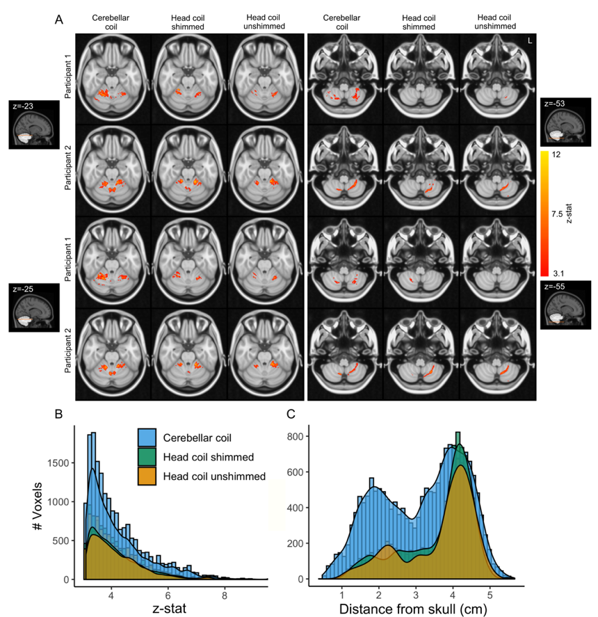
Figure 5: A, (Finger tapping > Rest) BOLD fMRI significant clusters (z>3.1) for 2 sample participants along axial slices at the height of the cerebellum, crossing the hand region in cerebellar lobule V (left) and cerebellar lobule VIII (right). B, Group Z-stat distribution for each coil. C, Barplots of active voxels at group level in relation to the distance from the skull.
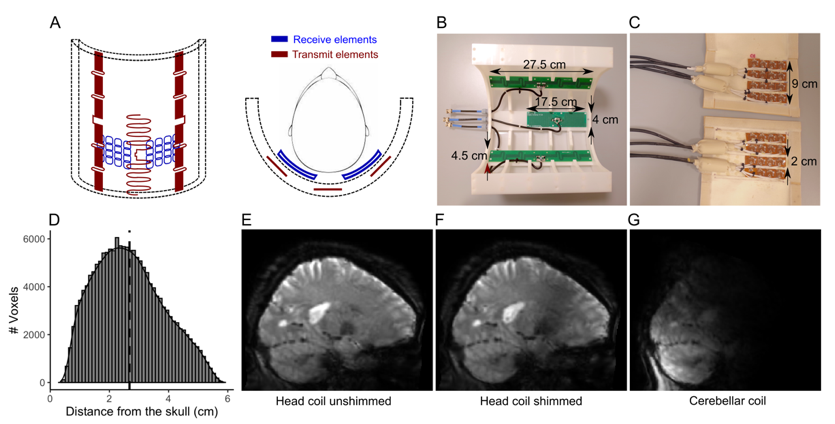
Figure 1: A, view of the cerebellar coil. Red, transmit elements. Blue, receive elements. B, The transmit array (housing partially removed). C, The receive array (open covers). D, Distribution of voxels in the cerebellum with respect to distance from the skull (median distance=2.58cm (dotted line)). E-G, 3D-EPI slices. E, Head coil without B1 shimming. F, Head coil after B1 shimming. G, Cerebellar coil. Note the reduced deeper sensitivity deeper due to the small total surface area.