Wahidul Alam1, Rushdi Zahid Rusho1, Scott Reineke2, Madavan Raja2, Stanley Kruger3, Joseph M. Reinhardt1, Junjie Liu4, Douglas Van Daele5, and Sajan Goud Lingala1,2
1Roy J Carver Department of Biomedical Engineering, University of Iowa, iowa city, IA, United States, 2ScanMed LLC, Omaha, NE, United States, 3Department of Radiology, University of Iowa, iowa city, IA, United States, 4Department of Neurology, University of Iowa, iowa city, IA, United States, 5Department of Otolaryngology, University of Iowa, iowa city, IA, United States
1Roy J Carver Department of Biomedical Engineering, University of Iowa, iowa city, IA, United States, 2ScanMed LLC, Omaha, NE, United States, 3Department of Radiology, University of Iowa, iowa city, IA, United States, 4Department of Neurology, University of Iowa, iowa city, IA, United States, 5Department of Otolaryngology, University of Iowa, iowa city, IA, United States
We develop a novel custom airway coil to offer significant boost in signal sensitivity in several upper-airway regions. The coil exhibits robust parallel MRI performance up to R=4~5 fold 1-D under-sampling for static imaging, and highly accelerated dynamic imaging (up to R~27 fold).
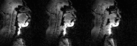
Fig.5 (animation): 2D concurrent multi-slice accelerated dynamic imaging of swallowing an ~10 ml bolus of pineapple juice. Non-Cartesian spiral under-sampling at ~27 fold acceleration level was combined with a sparse SENSE reconstruction scheme. The transport of the bolus is robustly captured in the three sagittal slices with adequate spatial resolution (2.4 mm2), and temporal resolution (17.1 ms).
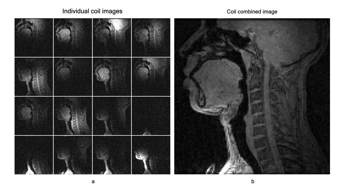
Fig.2: (a) Individual coil images from the 16 channel elements and (b) the R=1 SENSE coil combined image using the proposed airway coil. The coil offers high sensitivity in all upper-airway regions of interest (eg. lips, tongue, hard palate, soft palate, epiglottis, glottis).