Mohammed M. Albannay1, Charles McGrath1, Alexander Jaffray1, and Sebastian Kozerke1
1University and ETH Zurich, Institute for Biomedical Engineering, Zurich, Switzerland
1University and ETH Zurich, Institute for Biomedical Engineering, Zurich, Switzerland
SNR of thermal and hyperpolarized MRI is simulated based on first principles. Hyp. nuclei detection at low field strengths prolong T2* thus reduce readout bandwidth, leading to higher SNR compared to clinical field strengths. SNR gains from coil cooling are studied experimentally at 0.75T
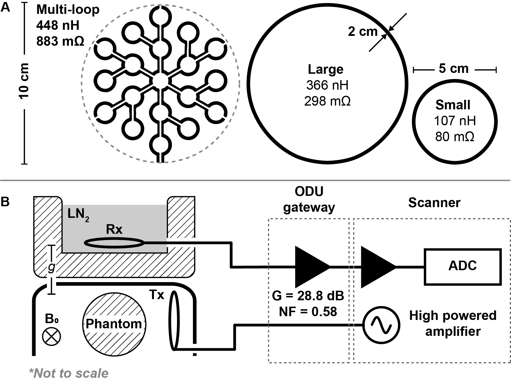
(A) Geometry of the three used receive coils with measured inductance and resistance using a vector network analyser at 32 MHz. The multi-loop coil is printed on 1.6 mm FR4 (1 oz copper), while loop coils are made from $$$(\oslash2 mm)$$$ copper tubing. (B) The coils were hosted in a Styrofoam box and laid parallel to the box surface. Separation $$$g = 2$$$ cm. The phantom was positioned adjacent to a $$$(\oslash5 cm)$$$ transmit coil, under a plastic arc.
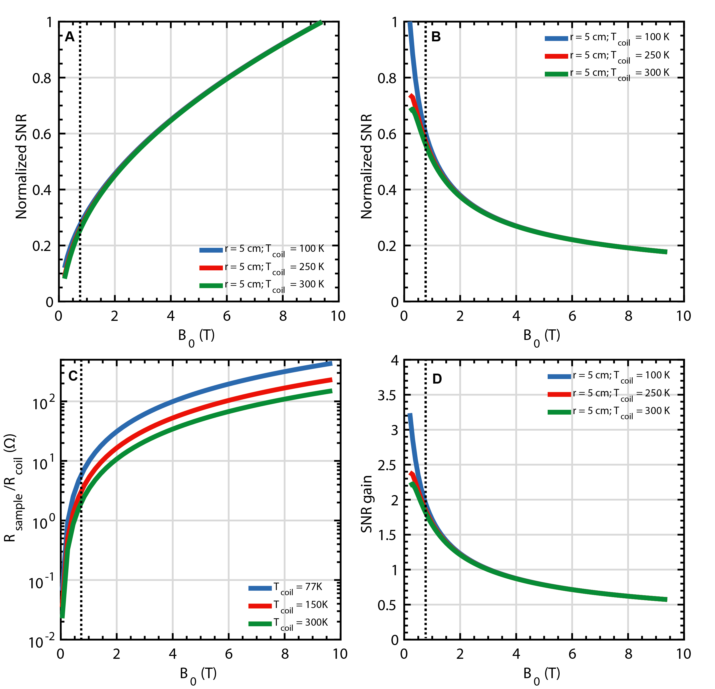
(A) Normalized simulated SNR for thermal and (B) hyperpolarized MRI as a function of field strength and coil temperature, (C) Ratio between equivalent sample and coil resistance. (D) Potential SNR gain relative to 300 K receiver coil at 3T for hyperpolarized MRI. All simulations consider a $$$\oslash$$$ 10 cm coil constructed from $$$\oslash $$$ 2 mm copper wire placed directly on the phantom. Inset dashed line indicating 0.75T.