Lena Nohava1,2, Andre Kuehne3, Elmar Laistler1, and Sigrun Roat1
1High Field MR Center, Center for Medical Physics and Biomedical Engineering, Medical University of Vienna, Vienna, Austria, 2BioMaps (Laboratoire d'Imagerie Biomédicale Multimodale Paris Saclay), Université Paris-Saclay, CEA, CNRS, Inserm, Orsay, France, 3MRI.TOOLS GmbH, Berlin, Germany
1High Field MR Center, Center for Medical Physics and Biomedical Engineering, Medical University of Vienna, Vienna, Austria, 2BioMaps (Laboratoire d'Imagerie Biomédicale Multimodale Paris Saclay), Université Paris-Saclay, CEA, CNRS, Inserm, Orsay, France, 3MRI.TOOLS GmbH, Berlin, Germany
For 7T MRI, 10 cm double-gap coaxial coil design operated at its self-resonance is advantageous for increased Tx efficiency and slightly lower SAR. Orientation independence of the coil’s B1-field is of importance for free positioning of flexible wearable arrays.
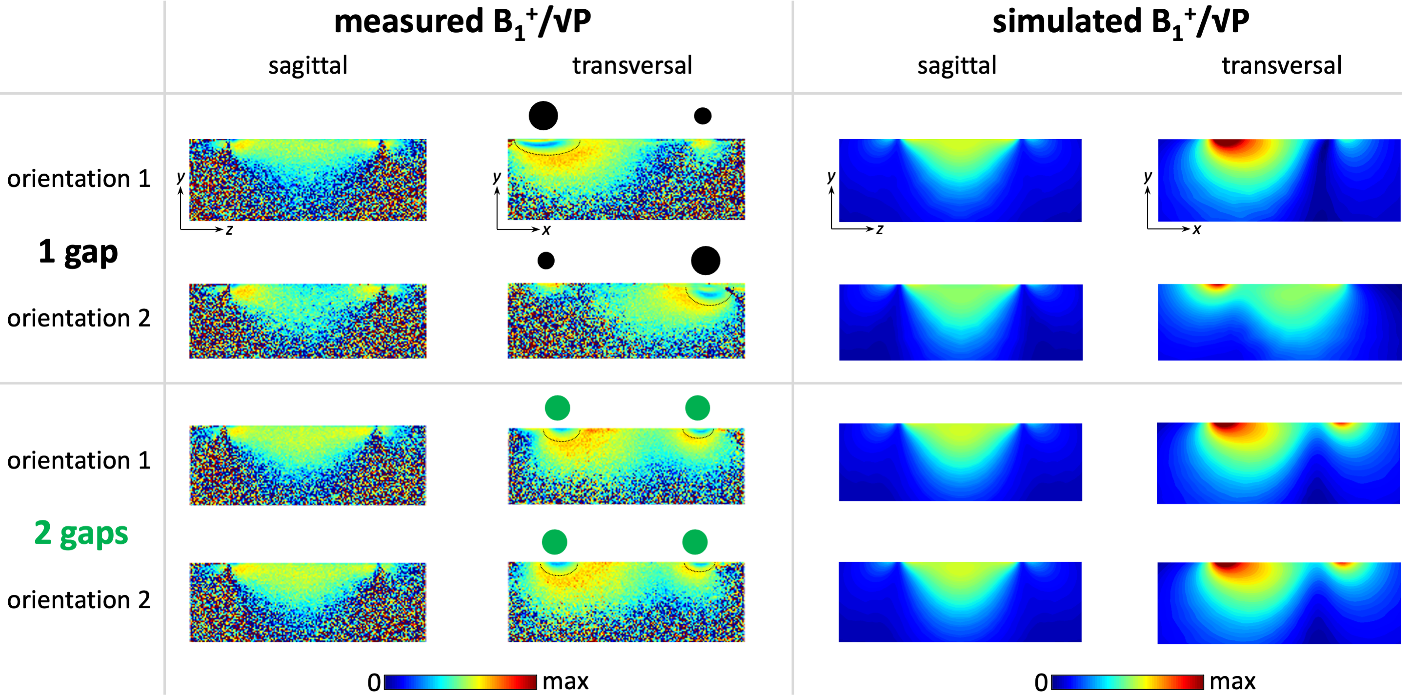
Figure 4: Measured and simulated B1+/√P maps in the central sagittal and transversal slice (FOV=256x256mm2, 0.5x0.5mm2 in-plane resolution, 1.5mm slice thickness, Vref=150V). Different orientations are indicated by black or green dots: their location corresponds to the coil conductor position and their size indicates the oCo current density (inhomogeneous for the 1G, homogeneous for the 2G coil). Thin black lines in transversal measured maps mark the boundaries of regions where the flip angle was higher than the dynamic range of the mapping technique.
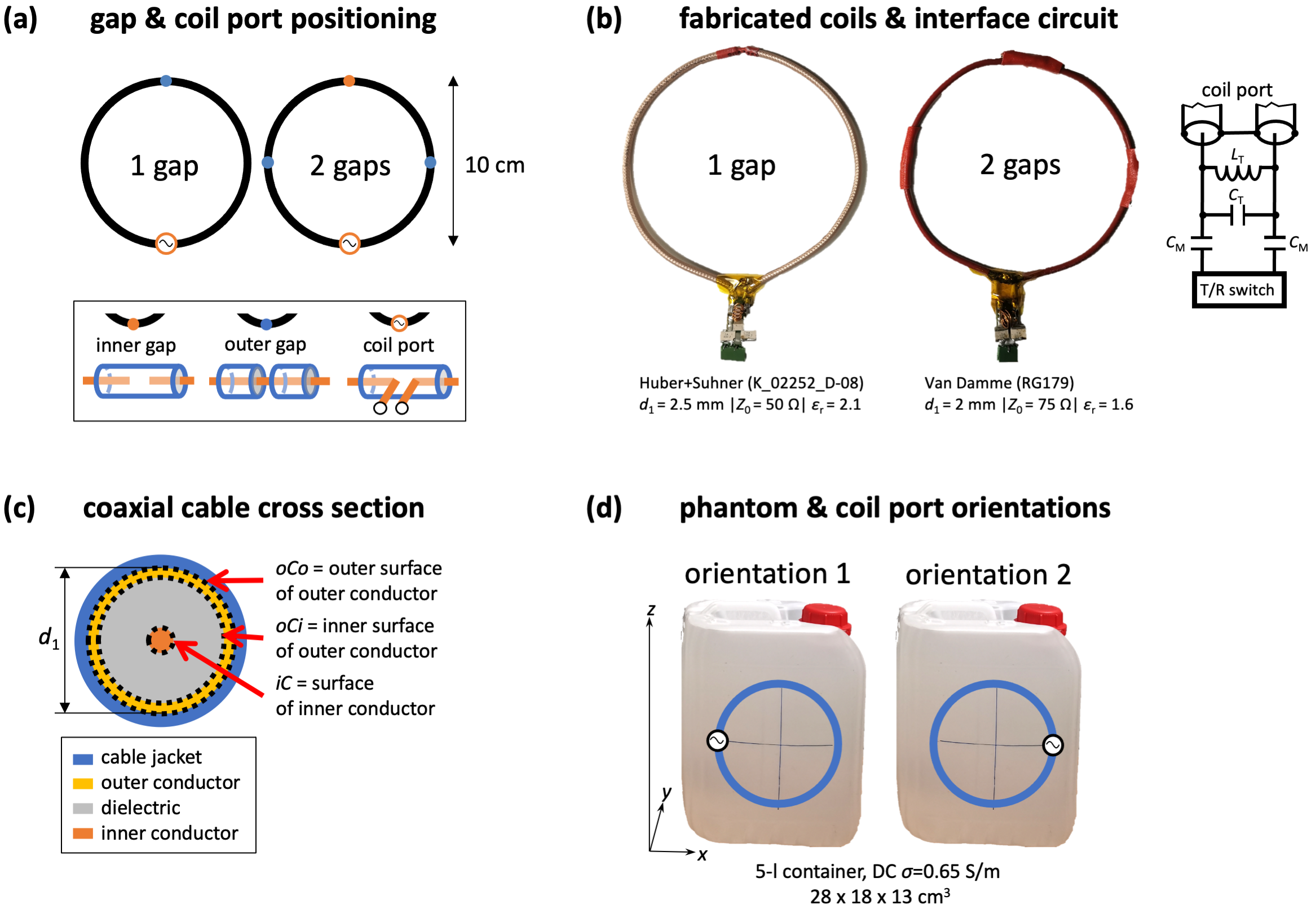
Figure 1: (a) Schematic showing gap positioning, (b) photographs of fabricated coils and interface circuit, (c) coaxial cable cross section, (d) phantom photo and investigated coil orientations.