Yun-Jeong Stickle1, Clyve Konrad Follante1, Mark Giancola1, David Anderson1, Fraser Robb1, Thomas Stickle1, Robert Stormont2, Holly Blahnik2, Ho-Joon Lee3, Young Han Lee4, and Darryl B. Sneag5
1GE Healthcare Coils, Aurora, OH, United States, 2GE Healthcare, Waukesha, WI, United States, 3Haeundae Paik Hospital, Busan, Korea, Republic of, 4Severance hospital, Yonsei University, Seoul, Korea, Republic of, 5Hospital for Special Surgery, New York, NY, United States
1GE Healthcare Coils, Aurora, OH, United States, 2GE Healthcare, Waukesha, WI, United States, 3Haeundae Paik Hospital, Busan, Korea, Republic of, 4Severance hospital, Yonsei University, Seoul, Korea, Republic of, 5Hospital for Special Surgery, New York, NY, United States
This study shows results for two different universally
sized AIR (Adaptive Imaging Receive) neck/cervical spine coils combined with a 48-Channel head coil to
provide higher SNR and improved acceleration compared to a conventional coil.

Fig. 1. Universally sized AIR 64-Channel
phased array head neck coils for carotid, head/neck and cervical spine MRI (a) Prototype1 16-Channel neck/cervical spine
anterior setup coil assembly with 48-Channel head coil (b) Prototype2
16-Channel neck/cervical spine posterior setup coil assembly with 48-Channel
head coil
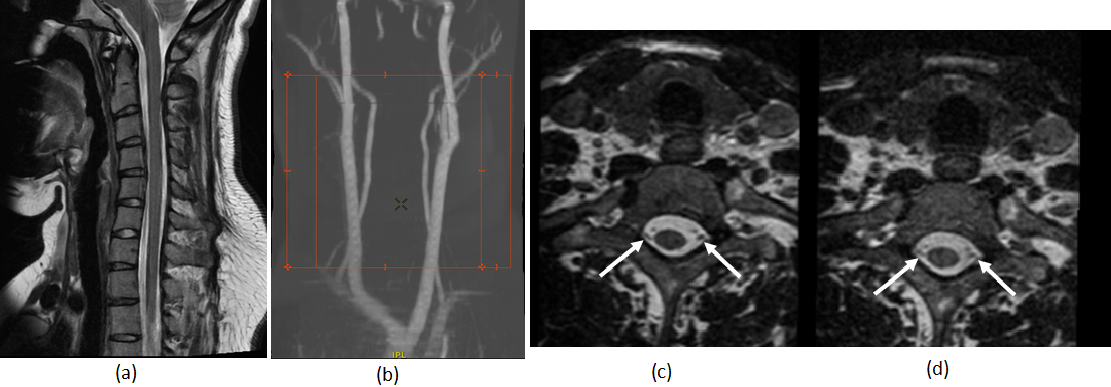
Fig. 5. (a) Sagittal T2,
20 cm FOV, 2mm slice thickness image and (b) MRA images obtained with the
protoytpe1 16-Channel neck/cervical spine coil with 48-Channel head coil. Axial
reformatted images of the cervical spine from a 3-D CUBE T2-weighted FSE sequence
(0.7mm isotropic) demonstrates improved SNR with the (c) protoytpe1 16-Channel
neck/cervical spine coil with 48-Channel head coil compared to the (d)
conventional coil. Note improved visualization of the intrathecal nerve
rootlets (arrows) with the prototype1 coil.