Bijaya Thapa1,2, Bernhard Strasser1,2, Xianqi Li1,2, Jason Stockman1,2, Azma Mareyam1, Boris Keil3, Zhe Wang4, Stefan Carp1,2, Yulin V. Chang4, Lawrence Wald1,2, Philipp Hoecht Hoecht5, and Ovidiu Andronesi1,2
1Dept. of Radiology, MGH, A. A. Martinos Center for Biomedical Imaging, Charlestown, MA, United States, 2Harvard Medical School, Boston, MA, United States, 3Mittelhessen University of Applied Science, Giessen, Germany, 4Siemens Medical Solutions USA, Charlestown, MA, United States, 5Siemens Healthcare, Erlangen, Germany
1Dept. of Radiology, MGH, A. A. Martinos Center for Biomedical Imaging, Charlestown, MA, United States, 2Harvard Medical School, Boston, MA, United States, 3Mittelhessen University of Applied Science, Giessen, Germany, 4Siemens Medical Solutions USA, Charlestown, MA, United States, 5Siemens Healthcare, Erlangen, Germany
An ERETIC method was integrated into 3D MRSI through the hardware and software for quantitative brain metabolite imaging and compared its result with the conventional internal water reference method. Bland-Altman plot plotted to compare them which shows good agreement between these methods.
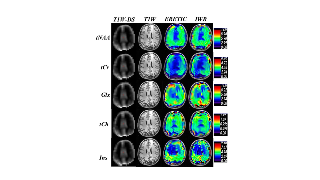
Fig3. Absolute
Metabolite concentration map in mMol unit obtained from ERETIC (3rd column)
and IWR (4th column) methods corresponding to the anatomical
MEMPRAGE image (2nd column) and MEMPRAG image down-sampled
to MRSI size (1st column ).
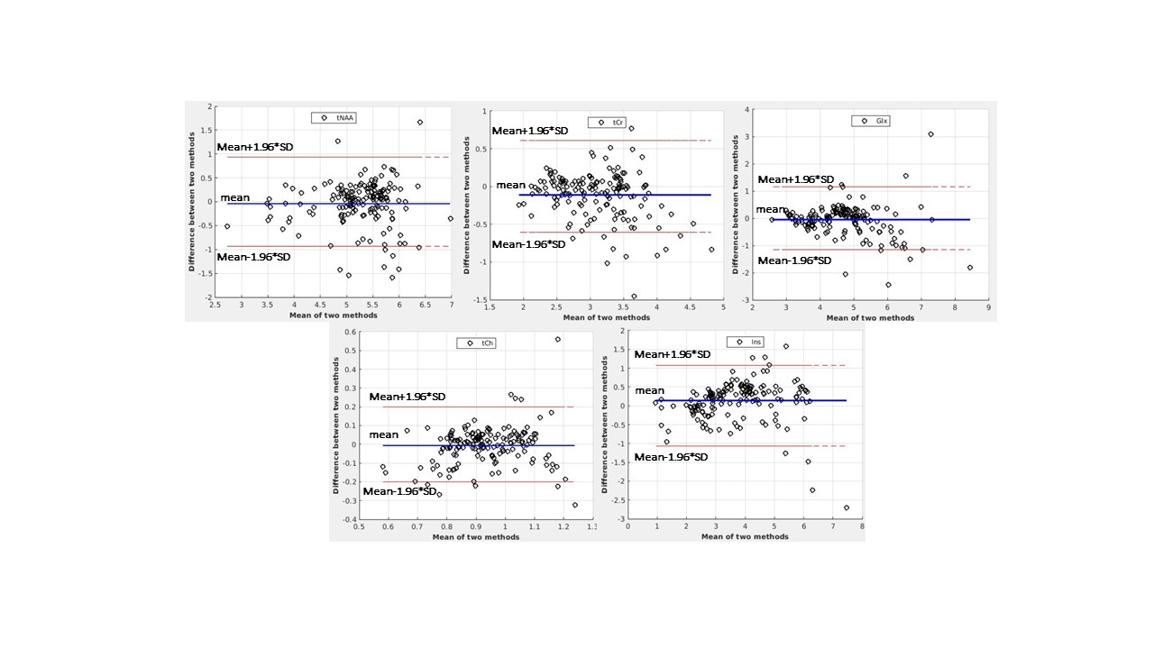
Fig4. Bland-Altman plots corresponding to the
metabolite concentration map shown in Fig.3, which indicates the agreement
between IWR and ERETIC methods.