Tijl van der Velden1, Mark Gosselink1, Ingmar Voogt2, Martijn Froeling1, Hans Hoogduin1, Dennis Klomp1, Bart Steensma1, and Alexander Raaijmakers1,3
1UMC Utrecht, Utrecht, Netherlands, 2Wavetronica, Utrecht, Netherlands, 3Biomedical Engineering, Eindhoven University of Technology, Eindhoven, Netherlands
1UMC Utrecht, Utrecht, Netherlands, 2Wavetronica, Utrecht, Netherlands, 3Biomedical Engineering, Eindhoven University of Technology, Eindhoven, Netherlands
An parallel imaging acceleration of a least a factor of 3 is
feasible with a 72-channel body array at 7T.
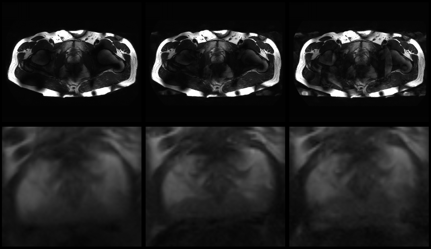
T2w
images with SENSE 1 (left), 2 (center) and 4 (right). Bottom row shows
magnification of the prostate. From an acceleration of 4 small artefacts are
observed, as well as the expected reduction in SNR.
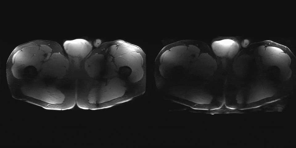
3D
T1w acquisition of the lower body. Left, no SENSE acceleration, right: 3x3
SENSE acceleration. Artefacts are predominately in the peripheral region of the
body.