El-Sayed H Ibrahim1, Xiaolong Wang1, and Bo Wang1
1Medical College of Wisconsin, Milwaukee, WI, United States
1Medical College of Wisconsin, Milwaukee, WI, United States
This
study showed that the developed graft has biomechanical characteristics similar
to those in native aorta and normal rat, which may represent a potential avenue to construct a
flexible vascular graft that matches the patient-specific dimension.
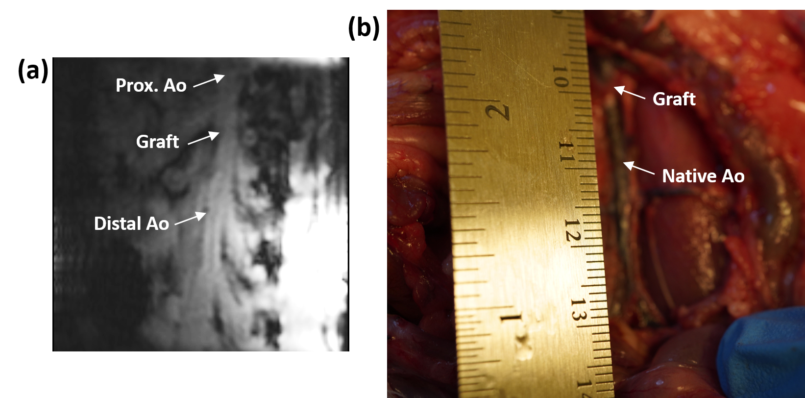
Figure 1. Coronal image showing native aorta and graft region, as well as
corresponding picture. The figure also shows locations where cross-sectional
phase-encoding images were acquired.
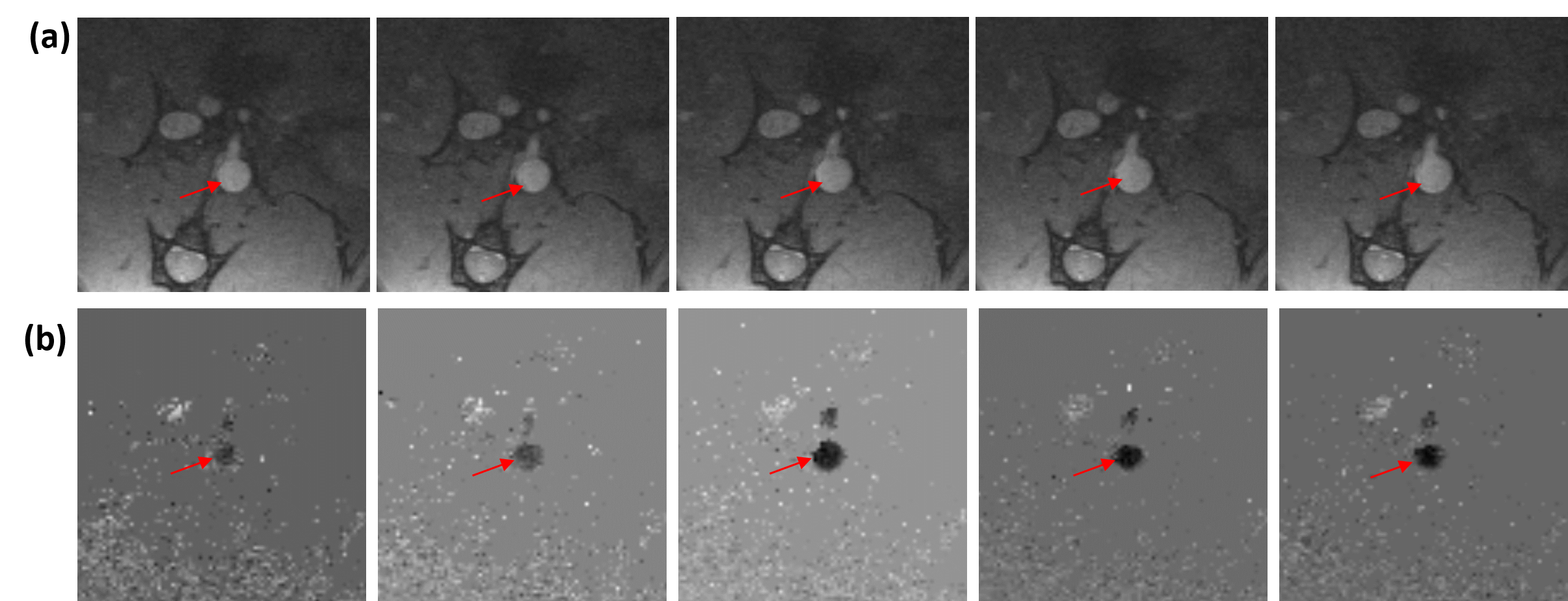
Figure 2. (a) Magnitude images showing a cross-sectional area in the graft region
(arrows), which grows with time during systole. (b) Velocity-encoded
phase-contrast images showing a cross-sectional area in the graft region
(arrows), where flow increases (blacker region) during early systole.