El-Sayed H Ibrahim1, Jamasp Azarnoosh2, Arash Hassankiadeh1, Pierre Croisille3, Jadranka Stojanovska4, and John LaDisa2
1Medical College of Wisconsin, Milwaukee, WI, United States, 2Marquette University, Milwaukee, WI, United States, 3Jean-Monnet University, Lyon, France, 4University of Michigan, Ann Arbor, MI, United States
1Medical College of Wisconsin, Milwaukee, WI, United States, 2Marquette University, Milwaukee, WI, United States, 3Jean-Monnet University, Lyon, France, 4University of Michigan, Ann Arbor, MI, United States
Cardiovascular
parameters can normalize in treated CoA compared to untreated cases. Myocardial
strain and aortic blood velocity are sensitive parameters for differentiating
between treated and untreated CoA as well as the severity of coarctation.
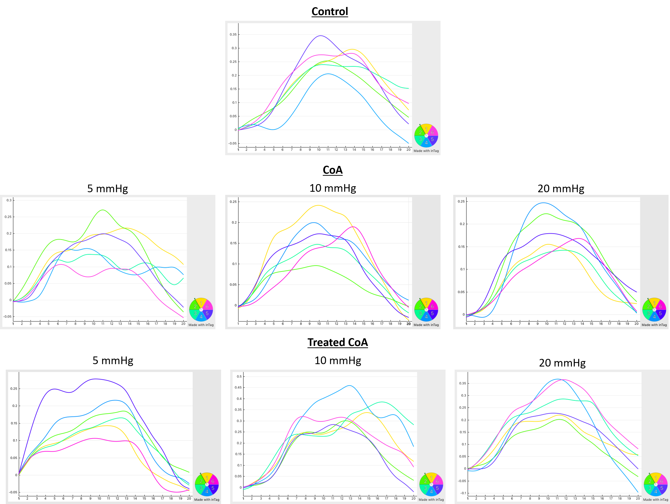
Figure 1. Strain curves in different rabbit models.
Circumferential strain curves throughout the cardiac cycle in different
segments of mid-ventricular short-axis slices. Strain is shown positive as
peripheral gating was used with peak systole during first timeframes. Note
decreased strain values with increased degree of coarctation. Strain values are
larger than those in untreated CoA (scale is different for different panels).
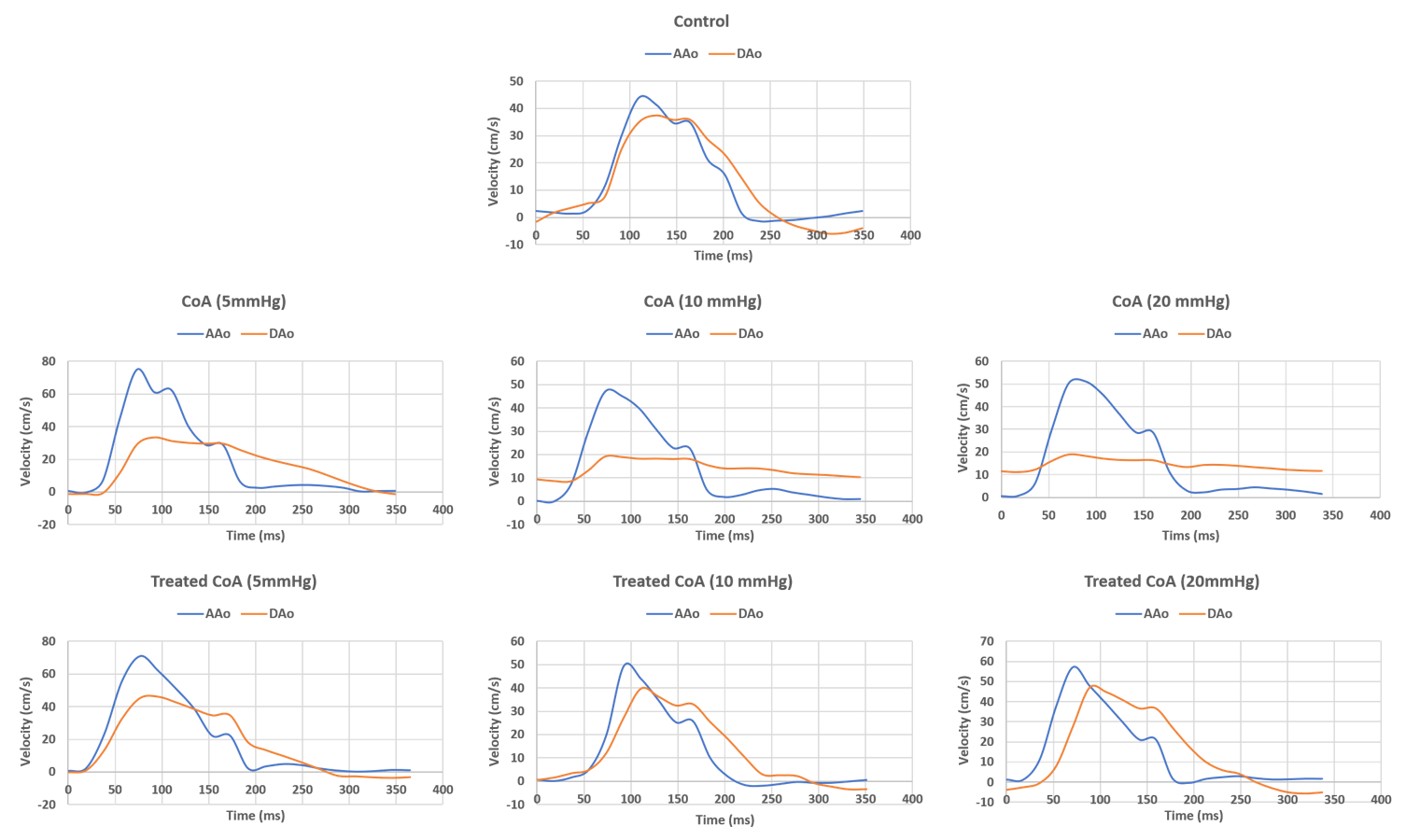
Figure 2. Velocity curves in different rabbit models. Mean velocity
throughout the cardiac cycle in ascending aorta (AAo) and proximal descending
aorta (DAo) in different rabbit models of CoA. The ratio between maximum
velocity in AAo to that in DAo was slightly >1 in control and treated CoA
compared to untreated CoA, where the ratio was always >2 in the latter.