Tianyi Zhou1, Liam Timms1, Valur Olafsson2, Fred Bidmead2, Vishala Mishra3, Ravi T Seethamraju4, Mukesh Harisinghani3, and Srinivas Sridhar1
1Department of Physics, Northeastern University, Boston, MA, United States, 2Northeastern University Biomedical Imaging Center, Boston, MA, United States, 3Department of Radiology, Massachusetts General Hospital, Boston, MA, United States, 4Siemens Medical Solutions, Boston, MA, United States
1Department of Physics, Northeastern University, Boston, MA, United States, 2Northeastern University Biomedical Imaging Center, Boston, MA, United States, 3Department of Radiology, Massachusetts General Hospital, Boston, MA, United States, 4Siemens Medical Solutions, Boston, MA, United States
A
contrast-enhanced MRA protocol with Ultrashort Time-to-Echo was optimized
on phantoms covering a range of ferumoxytol concentrations for abdominal
imaging.
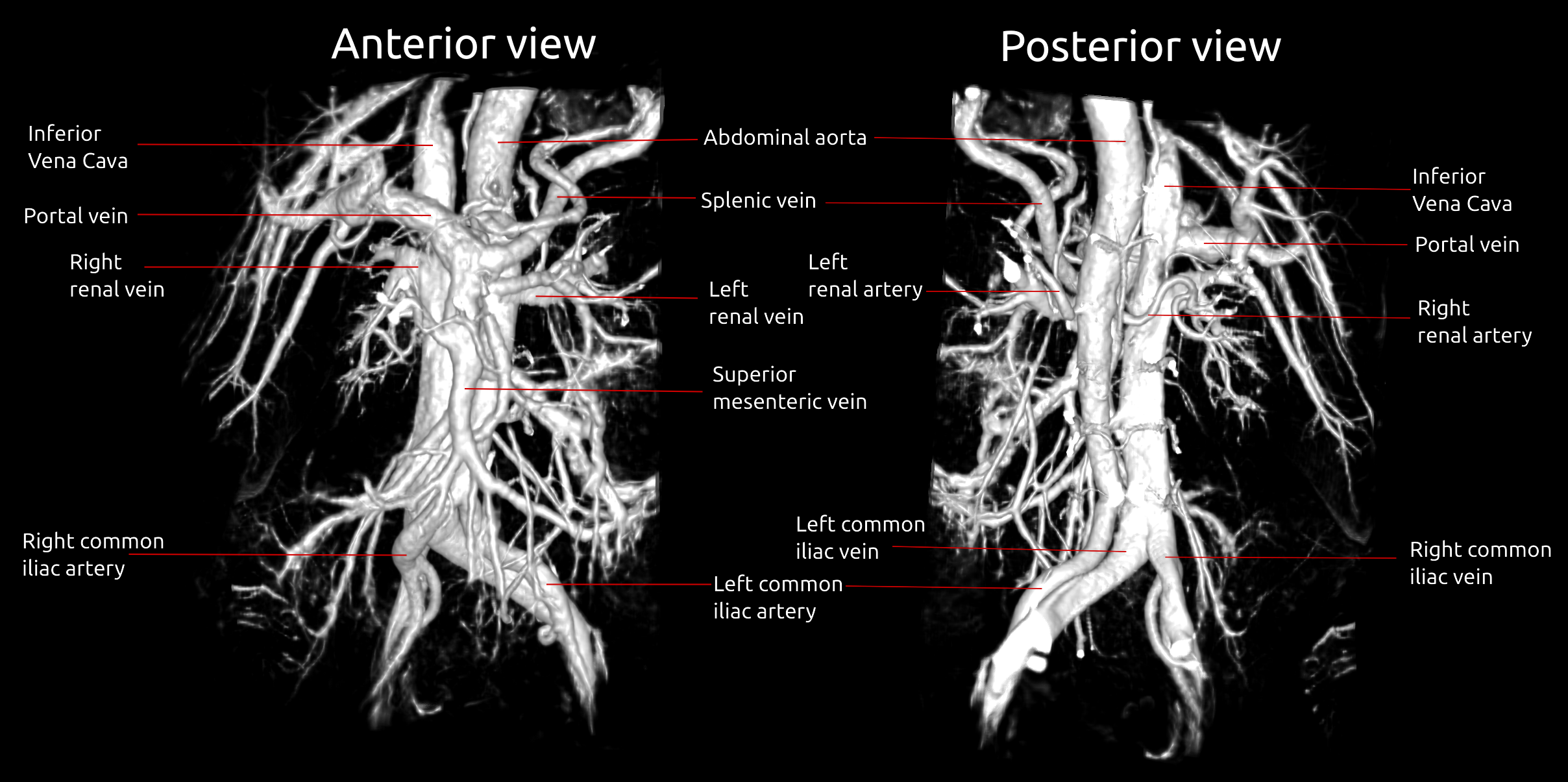
3D
rendering of the QUTE-CE image capturing the abdominal vascular anatomy shown
in anterior and posterior view.
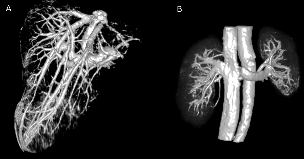
3D
rendering of the vasculature of the liver (A) and the kidneys (B) cropped from
the same QUTE-CE image shown in Figure 2.