Hao Li1, Martin John Graves1,2, Nadeem Shaida2, Akash Prashar2, David John Lomas1,2, and Andrew Nicholas Priest1,2
1Department of Radiology, University of Cambridge, Cambridge, United Kingdom, 2Department of Radiology, Addenbrooke’s Hospital, Cambridge, United Kingdom
1Department of Radiology, University of Cambridge, Cambridge, United Kingdom, 2Department of Radiology, Addenbrooke’s Hospital, Cambridge, United Kingdom
A time-efficient
high-resolution 3D fresh-blood imaging
technique based on compressed sensing is developed, which improved overall
vessel sharpness and small vessel depiction for peripheral angiography.
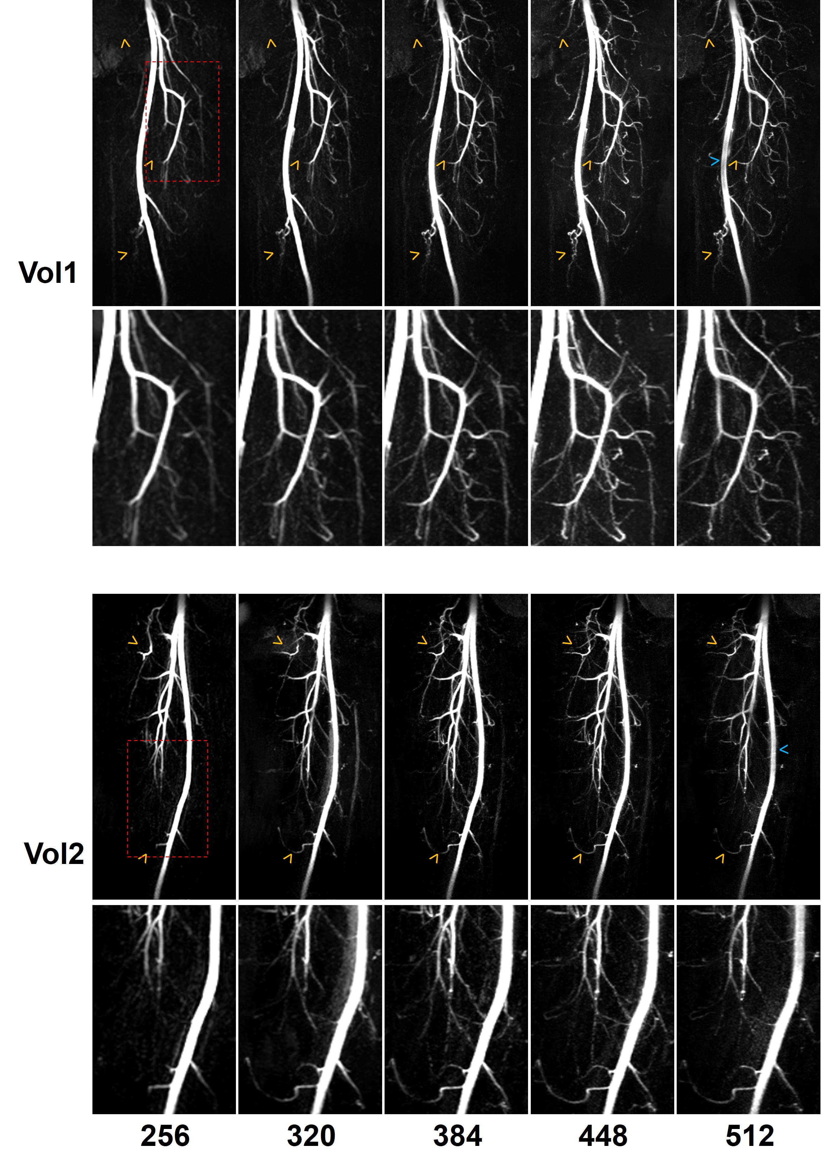
Figure 3. Example MIP images (upper
rows) and their zoomed-in views (lower rows, from the red dashed boxes) with
different resolutions from two healthy volunteers. The yellow arrowheads denote
the improved depiction of small arterial branches, and the blue arrowheads
denote the slight signal loss in the large artery on the image with very high
resolution (512×512).
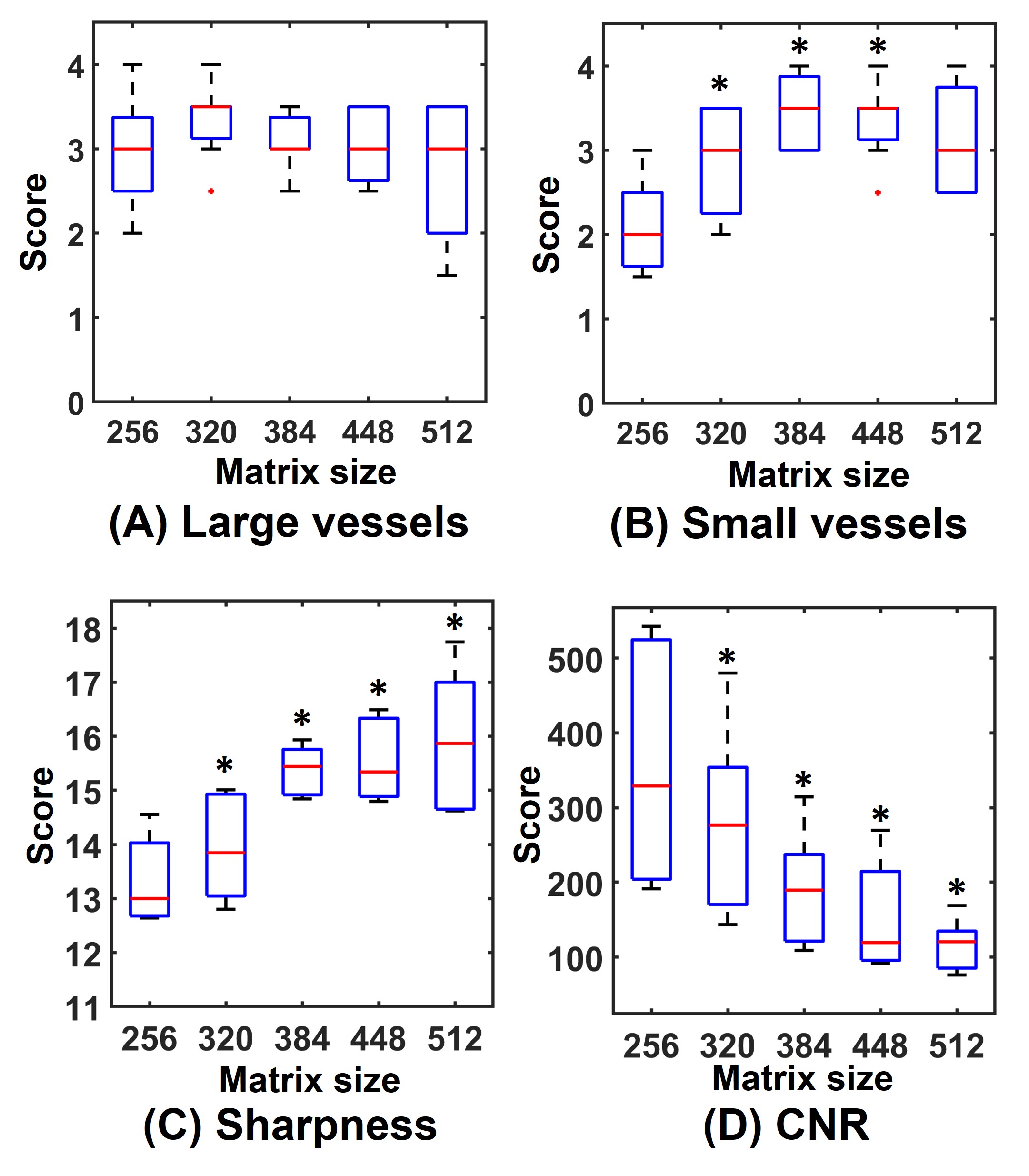
Figure 4. Box-plots of quality scores of
FBI images with different resolutions: (A) subjective evaluation of large
vessel depictions, (B) subjective evaluation of small vessel delineations, (C) objective
evaluation of sharpness and (D) objective evaluation of CNR of
artery-to-background. * denotes statistical significance between the
high-resolution images and the standard-resolution images (256×256). (Wilcoxon
signed-rank tests for A and B; paired t-tests
for C and D. P<0.05).