Akos Varga-Szemes1, Pascale Aouad2, U. Joseph Schoepf1, Tilman Emrich1, Basel Yacoub1, Thomas M Todoran3, Ioannis Koktzoglou4, and Robert R Edelman4
1Department of Radiology and Radiological Science, Medical University of South Carolina, Charleston, SC, United States, 2Northwestern University, Chicago, IL, United States, 3Department of Medicine, Medical University of South Carolina, Charleston, SC, United States, 4Department of Radiology, Northshore University HealthSystem, Evanston, IL, United States
1Department of Radiology and Radiological Science, Medical University of South Carolina, Charleston, SC, United States, 2Northwestern University, Chicago, IL, United States, 3Department of Medicine, Medical University of South Carolina, Charleston, SC, United States, 4Department of Radiology, Northshore University HealthSystem, Evanston, IL, United States
Overall
image quality and accuracy were not different between tsSOS-QISS and 2D-QISS,
indicating that 3D tsSOS-QISS provides similar diagnostic value in patients
with PAD to a standard commercially available 2D-QISS technique.
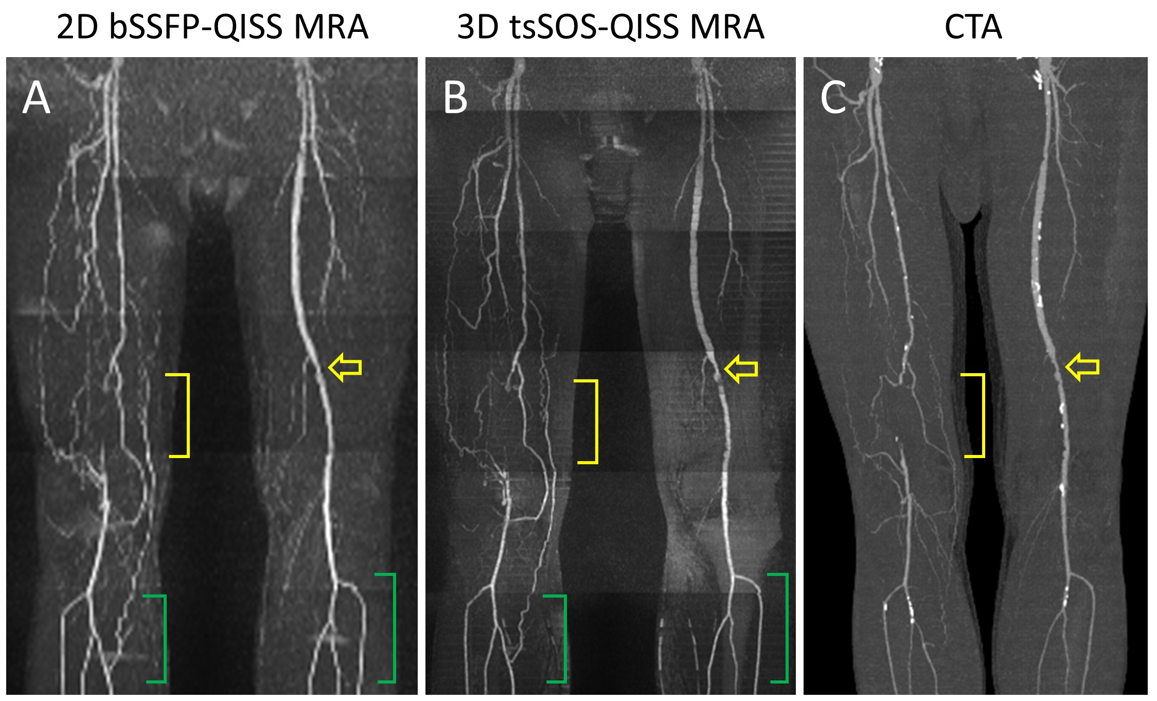
Figure 1 – 69-year-old man with PAD. Corresponding standard 2D-QISS MRA (A), 3D tsSOS-QISS MRA (B), and CTA (C) are shown. Total occlusion in the right distal superficial
femoral artery and popliteal artery (yellow
brackets) and significant stenosis in the left superficial femoral artery (yellow open arrows) are visualized. Stair-step
artifacts can be seen on 2D-QISS (A, green
brackets), which are not present on the corresponding 3D tsSOS-QISS (B, green brackets).