Kavya Sinha1, Christof Karmonik2, Alan B Lumsden1, and Trisha Roy1
1DeBakey Heart & Vascular Center, Houston Methodist Hospital, Houston, TX, United States, 2Translational Imaging Center, Houston Methodist Research Institute, Houston, TX, United States
1DeBakey Heart & Vascular Center, Houston Methodist Hospital, Houston, TX, United States, 2Translational Imaging Center, Houston Methodist Research Institute, Houston, TX, United States
Clinical high-field MRI protocol was established for the
visualization and quantification of PAD lesion components. Ex-vivo
imaging supported and confirmed the findings and
initial impression of the 7T images.
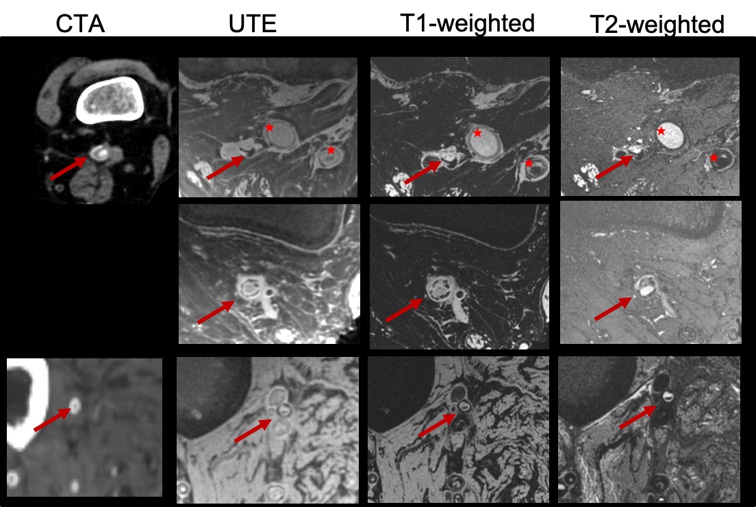
Figure 1: Multi-contrast images (CTA:
computed tomography angiography, UTE: ultra-short TE, T1-weighted, T2-weighted)
demonstrating excellent contrast between PAD lesion components in popliteal
(first and second row, red arrows) and peroneal artery (third row, red
arrows). Circumferential calcifications, as well as occlusions, can be readily appreciated due to the intrinsic high
spatial resolution achievable at the 7T scanner compared to the CTA. No CTA
available, 2nd row. (Red stars: Occluded PTFE grafts)
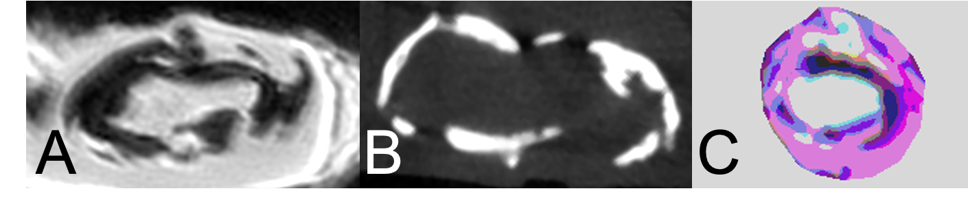
Figure 2: 3D UTE microMRI
(A) and corresponding microCT (B) image of the excised lesion
demonstrating good agreement between hypointense area on the micro MRI and the circumferential calcification
identified with the microCT.
Pseudo-color image (C) created from the 7T images illustrate the
differentiation power of the near in-vivo 7T to differentiate lesion components
also in respect to the ex-vivo microCT.