Evan Norris1, Guenther Schneider2, Toshimasa Clark1, Miles Kirchin3, Gregory Wilson4, and Jeffrey Maki1,4
1Radiology, University of Colorado, Aurora, CO, United States, 2University Hospital of Saarland, Homburg, Germany, 3Bracco Imaging, Milan, Italy, 4University of Washington, Seattle, WA, United States
1Radiology, University of Colorado, Aurora, CO, United States, 2University Hospital of Saarland, Homburg, Germany, 3Bracco Imaging, Milan, Italy, 4University of Washington, Seattle, WA, United States
We validate that
the theoretical relationship between gadolinium-based contrast agent (GBCA) concentration and R1, R2* in first pass contrast enhanced MRA (CE-MRA) allows for accurate predictions of CE-MRA
signal intensity for any given blood concentration of three different GBCAs.
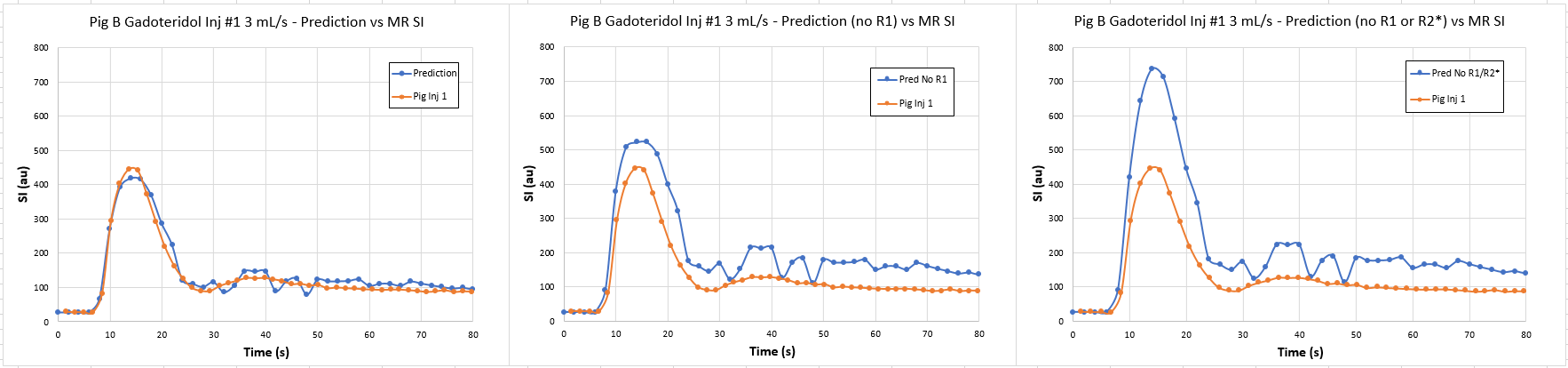
Figure
5: First gadoteridol injection (3 mL/s - left Figure 2) plots predicted (blue)
vs observed (orange) which integrates R1 and R2* effects
with excellent correlation between predicted and observed SI. The middle graph shows
the same predictive model but excludes R1 effects, with decreased
accuracy between predicted and observed SI. The right graph shows the same
predictive model without both R1 and R2* effects, which
is the least accurate construct. This is consistent across all agents and
injections.
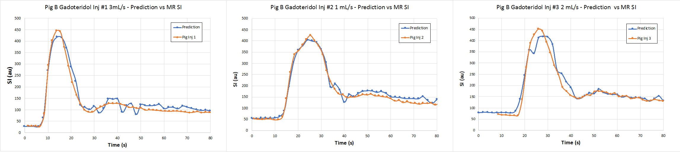
Figure
2: Three consecutive injections of gadoteridol at 3 (left), 1 (middle), and 2
(right) mL/s with predicted (blue) versus observed (orange) SI line plots. Relative
gain between observed and predictive SI was adjusted for the first injection
baseline, and held constant for the subsequent two injections. Blood R1 and R2* effects were
integrated into the model, with excellent correlation between predicted and
observed SI across all three injections.