Zechen Zhou1, Niranjan Balu2, Holger Eggers3, Peter Börnert3, Thomas S. Hatsukami2, and Chun Yuan2
1Philips Research North America, Cambridge, MA, United States, 2Vascular Imaging Lab, University of Washington, Seattle, WA, United States, 3Philips Research Hamburg, Hamburg, Germany
1Philips Research North America, Cambridge, MA, United States, 2Vascular Imaging Lab, University of Washington, Seattle, WA, United States, 3Philips Research Hamburg, Hamburg, Germany
The developed duel-echo
3D-MERGE with adiabatic flow suppression can achieve improved image quality for
large-coverage carotid vessel wall imaging. In addition, it can provide
quantitative fat fraction and field maps for potential plaque component analysis.
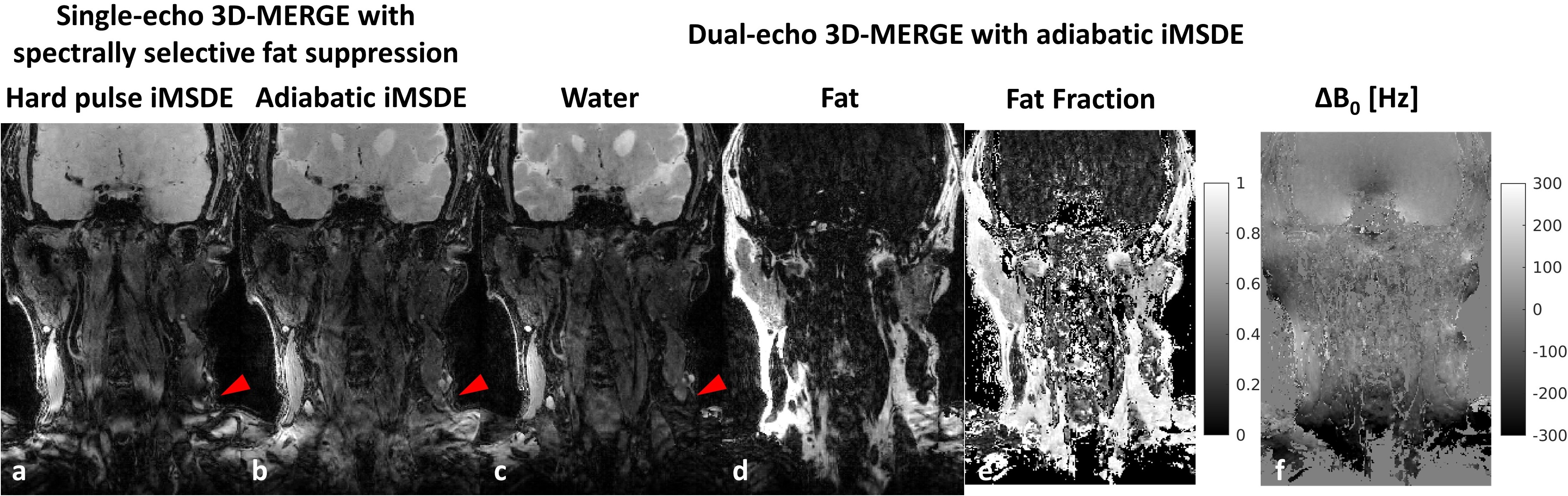
Figure
2: Comparison of different 3D-MERGE scans on a volunteer (coronal view). (a)
single-echo 3D-MERGE with composite hard pulse iMSDE and SPIR fat suppression.
(b) single-echo 3D-MERGE with adiabatic iMSDE and SPIR fat suppression. Water
(c) and fat (d) images as well as fat fraction (e) and field (f) maps are
decomposed from dual-echo 3D-MERGE with adiabatic iMSDE. Note the signal
intensity uniformity and fat suppression difference in those red arrow pointed
regions in (a)-(c).
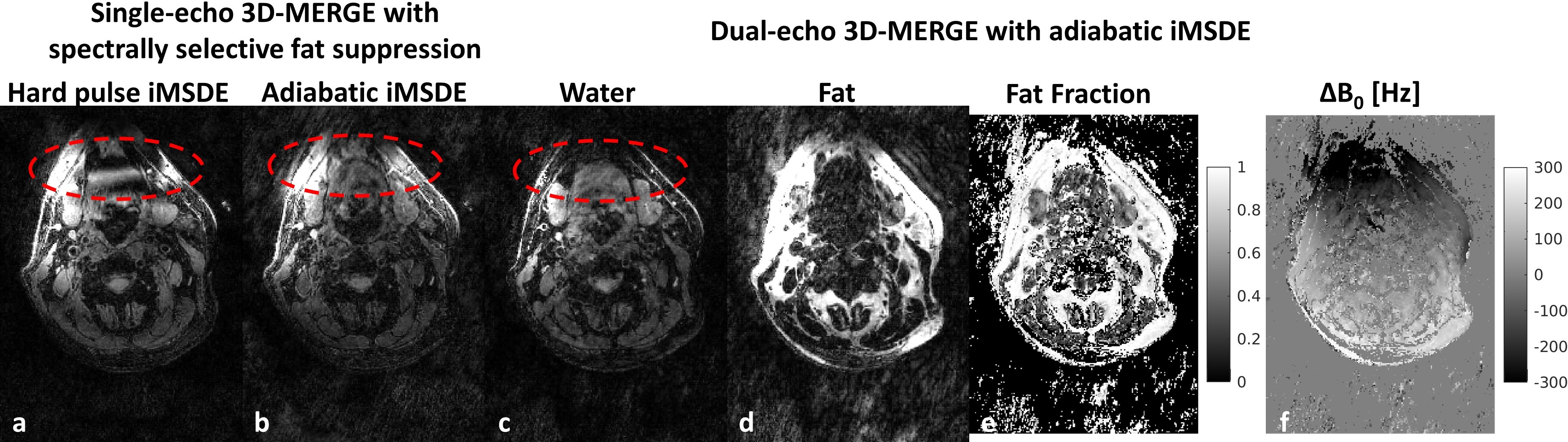
Figure 3: Comparison
of different 3D-MERGE scans on a volunteer (axial view). (a) single-echo
3D-MERGE with composite hard pulse iMSDE and SPIR fat suppression. (b)
single-echo 3D-MERGE with adiabatic iMSDE and SPIR fat suppression. Water (c)
and fat (d) images as well as fat fraction (e) and field (f) maps are
decomposed from dual-echo 3D-MERGE with adiabatic iMSDE. Note the signal
intensity uniformity and fat suppression difference in those red circle regions
in (a)-(c).