Takashi Namiki1, Hiroshi Hamano1, Naoki Udo2, Inka Ristow3, Felicia-Marie von Düring3, Alexander Lenz3, Shuo Zhang4, and Masami Yoneyama1
1Philips Japan, Tokyo, Japan, 2Department of Radiological technology , Yuuai Medical Center, Okinawa, Japan, 3Department of Diagnostic and Interventional Radiology and Nuclear Medicine, University Hospital Hamburg-Eppendorf, Hamburg, Germany, 4Philips, Hamburg, Germany
1Philips Japan, Tokyo, Japan, 2Department of Radiological technology , Yuuai Medical Center, Okinawa, Japan, 3Department of Diagnostic and Interventional Radiology and Nuclear Medicine, University Hospital Hamburg-Eppendorf, Hamburg, Germany, 4Philips, Hamburg, Germany
Free-breathing black-blood MRI of the main thoracoabdominal vessels
using MSDE-prepared radial imaging is possible without additional motion
compensation. A uniform blood suppression independent from the contrast agent
T1-shortening effect may permit assessment of wall abnormalities.
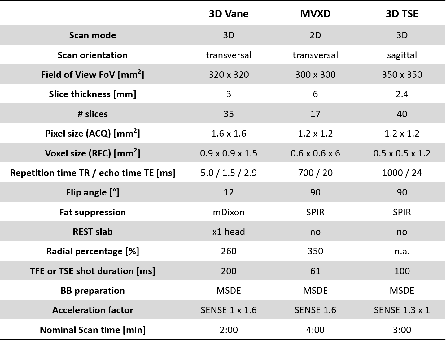
Table 1. Imaging
parameters of free-breathing MSDE-prepared 3D golden-angle radial
stack-of-stars (3D Vane-MSDE) and 2D TSE MultiVane XD (MVXD-MSDE)
used in this study. Conventional
Cartesian 3D TSE with MSDE-preparation and motion compensation was performed
for comparison (3D TSE-MSDE).
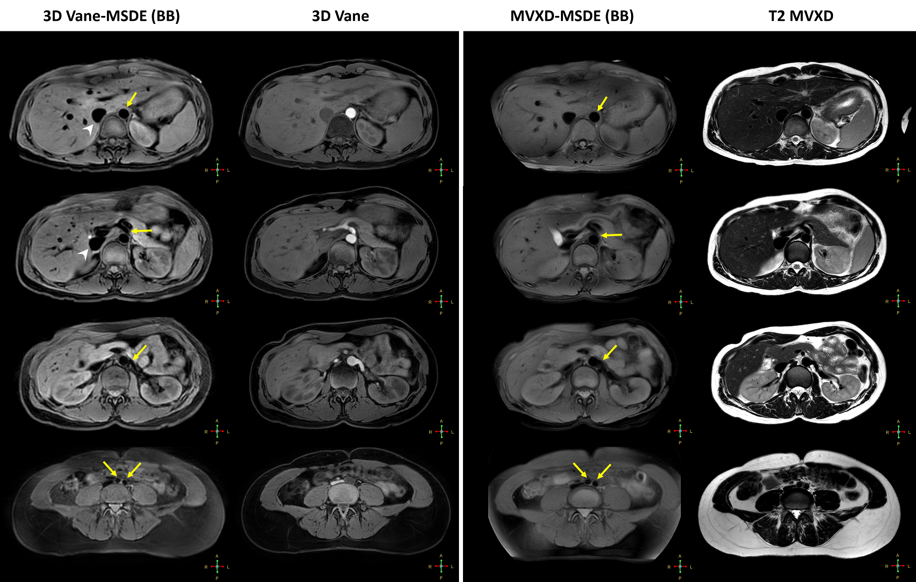
Figure 2. Free-breathing black-blood imaging with
proposed 3D Vane-MSDE and MVXD-MSDE. Homogeneous blood suppression was achieved by
MSDE preparation in both aorta (yellow
arrows) and inferior vena cava (arrow
heads). Images were selected at four different levels from top of the
kidney to aortic bifurcation in a healthy volunteer. Conventional bright-blood
3D Vane and T2 MVXD images at the corresponding slice locations were shown for
comparison.