Daichi Murayama1, Takayuki sakai1, Masami Yoneyama2, and Shigehiro Ochi3
1Radiology, Eastern Chiba Medical Center, Chiba, Japan, 2Philips Japan, Tokyo, Japan, 3Eastern Chiba Medical Center, Chiba, Japan
1Radiology, Eastern Chiba Medical Center, Chiba, Japan, 2Philips Japan, Tokyo, Japan, 3Eastern Chiba Medical Center, Chiba, Japan
Ultrashort- or zero-TE (uTE) MRA based Arterial Spin Labeling (ASL)has limitations because it requires koosh-ball sampling with whole brain coverage. we combined 3D radial sequences using stack-of-stars acquisition with uTE sampling to achieve fast imaging with focal slice coverages.
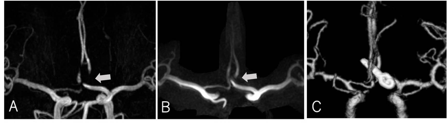
Fig.3 This 68-year-old woman had an anterior communicating artery aneurysm and was treated with surgical clipping. Maximum intensity projection (MIP) images obtained by TOF-MRA (A) and uTE-MRA (B). The volume rendering image reconstructed from computed tomography angiography (C).
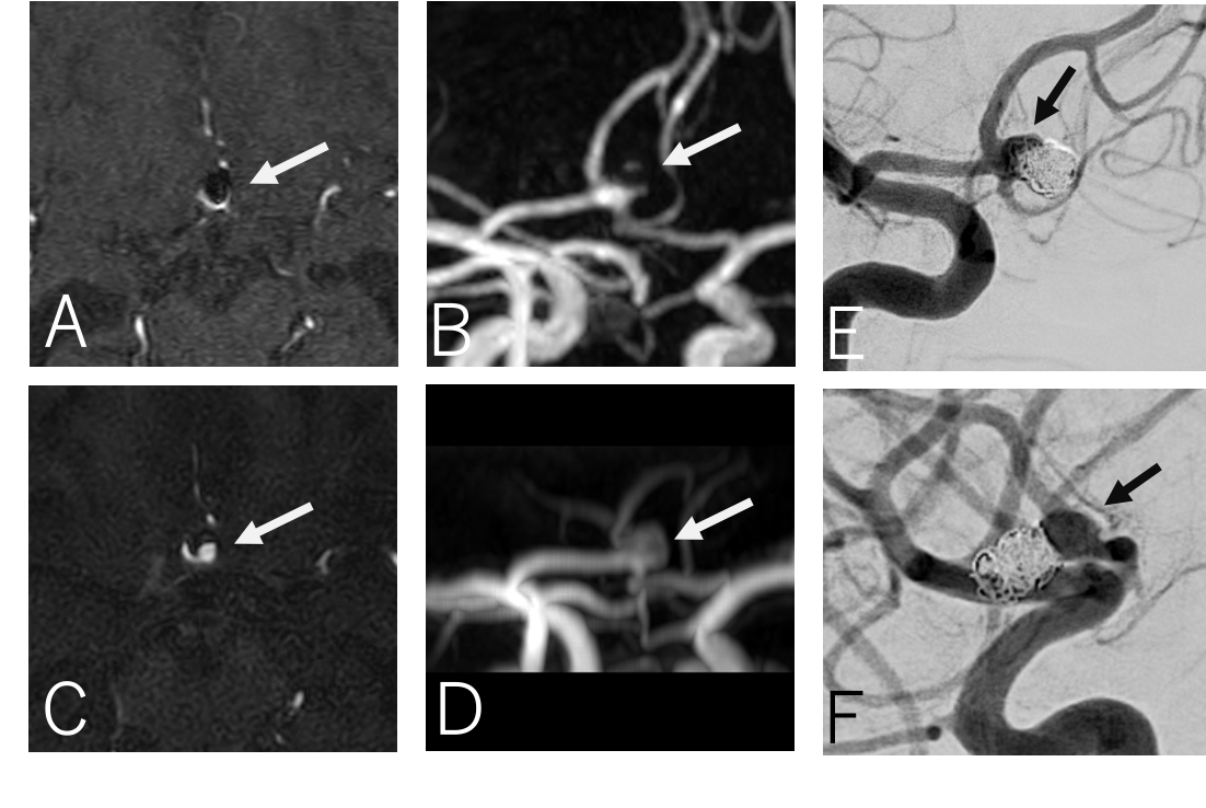
Fig.4 This 46-year-old woman had an anterior communicating artery aneurysm and was treated with coil embolization. Axial images and maximum intensity projection (MIP) images obtained by TOF-MRA (A, B) and uTE-MRA (C, D). Neck remnant was confirmed in follow-up MRA images obtained 6 months after coil embolization (arrows). DSA images were obtained in anteroposterior(E) lateral(F) views. Neck remnant was also observed in the DSA images taken after MRA imaging (arrows).